A Patient Presented With A Right Ankle Fracture
Onlines
Mar 13, 2025 · 7 min read
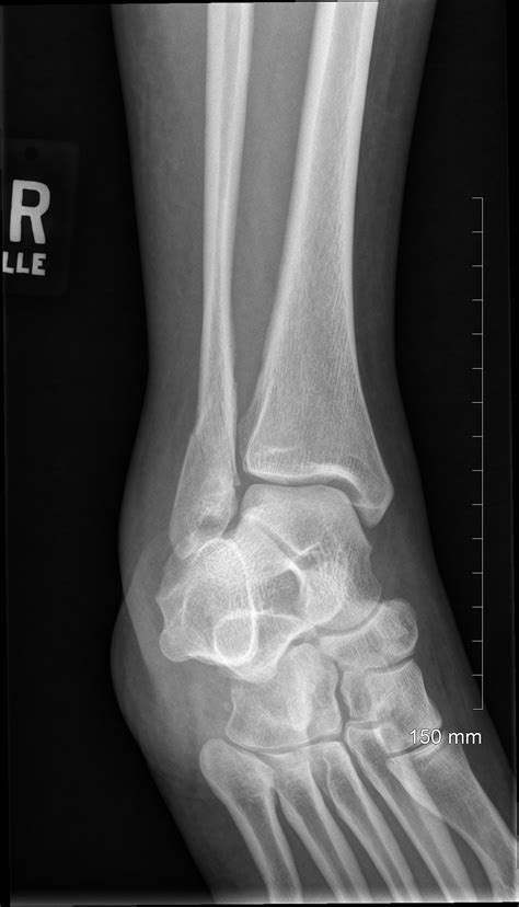
Table of Contents
A Patient Presented with a Right Ankle Fracture: A Comprehensive Overview
A patient presenting with a right ankle fracture requires a thorough and systematic approach encompassing initial assessment, diagnosis, treatment, and rehabilitation. Ankle fractures, encompassing breaks in one or more of the three bones forming the ankle joint (the tibia, fibula, and talus), represent a significant orthopedic challenge, demanding a nuanced understanding of the injury's complexity and potential long-term implications. This article delves into the various aspects of managing a patient with this specific presentation, emphasizing the importance of a multidisciplinary approach to ensure optimal patient outcomes.
Initial Assessment and Examination
The initial assessment of a patient with a suspected right ankle fracture begins with a comprehensive history taking and a detailed physical examination. The history should focus on the mechanism of injury, including the specifics of the event leading to the fracture. Understanding the force involved (high-energy versus low-energy trauma), the direction of the force, and any associated injuries provides crucial information for guiding the diagnostic process and subsequent treatment planning.
Key Aspects of the History:
- Mechanism of Injury: Was it a fall from height, a twisting injury during sports, a direct blow, or a motor vehicle accident? High-energy mechanisms are more likely to be associated with complex fractures and potential ligamentous injuries.
- Pain Characteristics: The location, severity, and type of pain (sharp, dull, aching) are essential for determining the extent and nature of the injury. Radiation of pain should also be noted.
- Associated Injuries: Ankle fractures often coexist with other injuries, such as fractures of the foot, lower leg, or even other body parts. Therefore, a thorough assessment of the entire lower extremity is critical.
- Past Medical History: Pre-existing conditions like osteoporosis, diabetes, or peripheral vascular disease can significantly impact healing and treatment options.
- Allergies and Medications: Documenting allergies and current medications is crucial for safe and effective management.
Physical Examination:
A thorough physical examination of the right ankle is paramount. This includes:
- Inspection: Observe for deformity, swelling, bruising (ecchymosis), and skin lacerations. Significant swelling and deformity strongly suggest a fracture.
- Palpation: Gently palpate the ankle joint for tenderness, crepitus (a grating sound indicating bone fragments rubbing together), and point tenderness over specific bony landmarks (malleoli, distal tibia, and fibula).
- Range of Motion: Assess active and passive range of motion. Significant limitation or pain with movement indicates significant injury.
- Neurovascular Assessment: Check the pulses (dorsalis pedis and posterior tibial), capillary refill, sensation, and motor function (dorsiflexion and plantarflexion of the foot) to detect any compromise to the neurovascular structures. This is crucial to identify potential complications like compartment syndrome.
- Stability Testing: Assess the stability of the ankle joint by gently stressing the joint in various directions. Significant instability indicates ligamentous injury in addition to the fracture.
Diagnostic Imaging
The physical examination provides valuable preliminary information, but definitive diagnosis requires imaging studies. X-rays are the primary imaging modality used to visualize ankle fractures. They typically include anteroposterior (AP), lateral, and mortise views. These views provide information on the location, type, and severity of the fracture.
Types of Ankle Fractures:
Ankle fractures are categorized based on the bones involved and the fracture pattern. Some common types include:
- Weber Classification: This system classifies fibula fractures based on their location relative to the distal tibiofibular syndesmosis (the joint connecting the tibia and fibula). Weber A fractures are below the syndesmosis, Weber B fractures are at the level of the syndesmosis, and Weber C fractures are above the syndesmosis.
- Lauge-Hansen Classification: This more complex system classifies ankle fractures based on the mechanism of injury and the fracture pattern. It incorporates the initial rotational and loading forces involved.
- Bimalleolar and Trimalleolar Fractures: These fractures involve two or three malleoli (the bony prominences of the ankle). Bimalleolar fractures involve both the medial and lateral malleoli, while trimalleolar fractures involve the medial, lateral, and posterior malleoli.
- Pilon Fractures: These are severe fractures involving the distal tibia, often resulting from high-energy trauma.
Additional imaging may be required in certain cases, including:
- Computed Tomography (CT) Scan: CT scans provide detailed three-dimensional images, which are useful for assessing complex fractures, identifying subtle fractures, and evaluating articular involvement. They are particularly helpful in planning surgical intervention.
- Magnetic Resonance Imaging (MRI): MRI is useful for evaluating soft tissue injuries, such as ligament sprains and cartilage damage, which often accompany ankle fractures. It is less commonly used for initial assessment but plays a crucial role in assessing the extent of ligamentous damage.
Treatment Options
Treatment of ankle fractures depends on several factors, including the type and severity of the fracture, the patient's age and overall health, and the presence of associated injuries. Treatment options range from non-surgical to surgical interventions.
Non-Surgical Management:
Non-surgical management, also known as conservative management, is suitable for certain stable ankle fractures, typically those with minimal displacement and good alignment. It usually involves:
- Immobilization: The ankle is immobilized using a cast, splint, or walking boot to maintain fracture alignment and prevent further displacement. The duration of immobilization depends on the fracture pattern and the healing process.
- Pain Management: Pain is managed with analgesics and nonsteroidal anti-inflammatory drugs (NSAIDs).
- Weight-Bearing Restrictions: Weight-bearing is restricted initially to protect the fracture and allow for proper healing. The degree of weight-bearing restriction is determined by the physician.
- Physical Therapy: Once the fracture has healed sufficiently, physical therapy is crucial to restore range of motion, strength, and functional mobility.
Surgical Management:
Surgical management is often necessary for unstable ankle fractures, those with significant displacement or malalignment, and fractures associated with ligamentous injuries. Surgical techniques vary depending on the fracture pattern and the surgeon's preference. Common surgical procedures include:
- Open Reduction and Internal Fixation (ORIF): This involves surgically exposing the fracture site, reducing (realigning) the fracture fragments, and stabilizing them with internal fixation devices such as screws, plates, or wires.
- External Fixation: External fixation involves attaching pins or screws to the bone fragments externally and connecting them to a frame outside the skin. This provides stabilization while allowing for early mobilization and wound care.
- Arthrodesis (Fusion): In severe cases where joint congruity cannot be restored or in cases of severe arthritis, arthrodesis may be necessary. This involves surgically fusing the ankle joint, resulting in a loss of motion but providing stability and pain relief.
Post-operative Care and Rehabilitation
Following surgical intervention, post-operative care is crucial for optimal healing and functional recovery. This involves:
- Wound Care: Meticulous wound care is essential to prevent infection.
- Pain Management: Pain is managed with analgesics as needed.
- Immobilization: The ankle may require further immobilization with a cast or splint, depending on the surgical procedure.
- Weight-Bearing Status: Weight-bearing restrictions vary depending on the type of surgery and the surgeon’s recommendations.
- Physical Therapy: Once the fracture has healed sufficiently, a comprehensive physical therapy program is critical. This includes range of motion exercises, strengthening exercises, and functional activities to regain normal ankle function.
Potential Complications
Several complications can arise following an ankle fracture, including:
- Nonunion: Failure of the fracture fragments to heal properly.
- Malunion: Healing of the fracture in a malaligned position.
- Infection: Infection at the fracture site or surgical incision.
- Compartment Syndrome: A condition characterized by increased pressure within the muscle compartments of the lower leg, potentially compromising blood supply and nerve function.
- Osteoarthritis: Development of osteoarthritis in the ankle joint due to post-traumatic changes.
- Chronic Pain: Persistent pain and dysfunction despite treatment.
- Complex Regional Pain Syndrome (CRPS): A chronic pain condition affecting the limb, often following injury.
Long-Term Outcomes and Follow-up Care
The long-term outcome after an ankle fracture depends on various factors, including the severity of the injury, the effectiveness of treatment, and the patient's compliance with rehabilitation. Regular follow-up visits are essential to monitor healing, assess for complications, and ensure optimal functional recovery. The patient should receive guidance on activity modification, appropriate footwear, and strategies for preventing future injuries. Return to pre-injury activities is gradual and depends on the extent of recovery.
This comprehensive overview of a patient presenting with a right ankle fracture highlights the complexity of this condition and underscores the importance of a thorough assessment, appropriate diagnostic imaging, timely and effective treatment, and a dedicated rehabilitation program. A multidisciplinary approach involving orthopedic surgeons, physical therapists, and other healthcare professionals is crucial to ensure the best possible outcomes for patients with ankle fractures. Early intervention and meticulous management can minimize complications and optimize functional recovery. The patient's active participation in the treatment process and compliance with the rehabilitation plan are paramount for achieving a favorable prognosis.
Latest Posts
Latest Posts
-
To Understand An Assertion Is To It
Mar 13, 2025
-
Drag The Tiles To The Correct Boxes
Mar 13, 2025
-
Unit 2 Understanding Functions Unit Test A Answer Key
Mar 13, 2025
-
How Does A Nursing Shortage Impact The Lpn Lvn
Mar 13, 2025
-
Statistics Unlocking The Power Of Data 3rd Edition Solutions Pdf
Mar 13, 2025
Related Post
Thank you for visiting our website which covers about A Patient Presented With A Right Ankle Fracture . We hope the information provided has been useful to you. Feel free to contact us if you have any questions or need further assistance. See you next time and don't miss to bookmark.
