An Infant With A History Of Tracheal Stenosis Quizlet
Onlines
Mar 13, 2025 · 6 min read
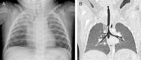
Table of Contents
An Infant with a History of Tracheal Stenosis: A Comprehensive Overview
Tracheal stenosis, a narrowing of the trachea (windpipe), is a serious condition that can significantly impact an infant's breathing and overall health. Understanding its various aspects, from diagnosis to management, is crucial for healthcare professionals. This article delves into the complexities of tracheal stenosis in infants, exploring its causes, symptoms, diagnostic methods, and treatment options. We will also examine potential complications and long-term implications for affected infants.
Understanding Tracheal Stenosis in Infants
Tracheal stenosis in infants refers to the narrowing of the trachea, the tube that carries air to and from the lungs. This narrowing can occur anywhere along the trachea, and its severity can range from mild to life-threatening. The condition can be congenital (present at birth) or acquired (develops after birth). The severity and location of the stenosis play a significant role in determining the symptoms and the necessary treatment approach.
Congenital Tracheal Stenosis: This form is often caused by abnormal development during fetal growth. Several factors can contribute, including genetic mutations, exposure to teratogens during pregnancy, and vascular rings (abnormal blood vessel development around the trachea).
Acquired Tracheal Stenosis: This type typically arises after birth due to various factors:
- Intubation-related injuries: Prolonged or traumatic intubation during surgery or intensive care can lead to tracheal damage and subsequent stenosis. This is a significant contributing factor in many cases.
- Tracheomalacia: This refers to the softening and collapse of the tracheal cartilage, which can lead to narrowing.
- Infections: Severe respiratory infections, such as whooping cough (pertussis) or bacterial tracheitis, can cause inflammation and scarring that narrow the airway.
- Trauma: External trauma to the trachea, such as injuries from blunt force or penetrating wounds, can cause stenosis.
- Tumors: Although less common, tumors within or near the trachea can cause compression and narrowing.
Symptoms of Tracheal Stenosis in Infants
The symptoms of tracheal stenosis can vary significantly depending on the severity and location of the narrowing. In severe cases, symptoms may be immediately apparent at birth, while in milder cases, they may not appear until later in infancy. Common symptoms include:
- Stridor: A high-pitched, wheezing sound during breathing, often heard most prominently during inhalation. This is a hallmark sign of tracheal stenosis.
- Respiratory distress: Difficulty breathing, characterized by rapid breathing, retractions (the chest wall sinking in during inhalation), and nasal flaring.
- Cough: A persistent, often harsh, cough.
- Cyanosis: A bluish discoloration of the skin and mucous membranes due to low blood oxygen levels. This is a serious sign indicating severe respiratory compromise.
- Apnea: Episodes of pausing in breathing.
- Recurrent respiratory infections: Infants with tracheal stenosis are more susceptible to respiratory infections due to the compromised airway.
- Poor weight gain: Difficulty breathing can make feeding and nutrient absorption challenging, leading to poor weight gain.
Severity of Symptoms: Mild stenosis may only present with subtle stridor during exertion, while severe stenosis can cause life-threatening respiratory distress.
Diagnosis of Tracheal Stenosis in Infants
Diagnosing tracheal stenosis involves a combination of clinical examination, imaging studies, and potentially bronchoscopy.
- Physical Examination: A thorough physical examination is the first step, focusing on assessing respiratory effort, auscultating for stridor and other abnormal breath sounds, and observing for signs of respiratory distress.
- Chest X-ray: This provides a preliminary view of the trachea and surrounding structures, helping to identify any gross abnormalities or associated conditions. However, it may not always clearly demonstrate the degree of stenosis.
- Computed Tomography (CT) Scan: A CT scan offers a more detailed three-dimensional image of the trachea, allowing for precise assessment of the location and extent of the stenosis. It’s crucial for detailed pre-operative planning.
- Magnetic Resonance Imaging (MRI): MRI provides excellent soft tissue detail and can be helpful in evaluating the surrounding structures and assessing for associated abnormalities, particularly vascular rings.
- Bronchoscopy: This minimally invasive procedure involves inserting a thin, flexible tube with a camera attached into the trachea to visualize the airway directly. It allows for precise measurement of the stenosis, assessment of its location and characteristics, and collection of tissue samples if necessary.
Treatment Options for Tracheal Stenosis in Infants
The treatment approach for tracheal stenosis depends on the severity of the stenosis, its location, and the presence of any associated conditions. Treatment options range from conservative management to surgical intervention.
- Conservative Management: In mild cases, conservative management may be sufficient. This may involve close monitoring, supportive care, and treating any associated respiratory infections promptly.
- Medical Management: Medical management may involve the use of bronchodilators to help relax the airway muscles and improve breathing. Steroids may also be used to reduce inflammation.
- Surgical Intervention: Surgical intervention is often necessary for moderate to severe stenosis. Several surgical techniques are available, including:
- Tracheoplasty: This involves surgically widening the narrowed section of the trachea. Different techniques exist depending on the nature and extent of the stenosis.
- Tracheal resection and anastomosis: This more involved procedure involves removing the severely stenotic section of the trachea and reconnecting the remaining ends. This requires careful planning and execution.
- Stent placement: A stent can be placed in the trachea to keep the airway open. However, stents can have potential complications, including migration, infection, and airway irritation.
Potential Complications and Long-Term Implications
Untreated or inadequately treated tracheal stenosis can lead to several serious complications, including:
- Respiratory failure: Severe respiratory compromise requiring mechanical ventilation.
- Pneumonia: Recurrent and severe respiratory infections, including pneumonia.
- Cor pulmonale: Right-sided heart failure due to chronic lung disease.
- Growth retardation: Difficulty breathing can interfere with feeding and nutrition, impacting growth.
- Death: In severe cases, tracheal stenosis can be life-threatening.
Long-term implications can vary depending on the severity of the stenosis and the effectiveness of the treatment. Some children may require ongoing monitoring and medical care, while others may lead relatively normal lives with minimal residual effects. The potential for recurrent stenosis or other long-term respiratory issues needs to be considered.
Prognosis and Follow-up Care
The prognosis for infants with tracheal stenosis depends heavily on several factors, including the severity of the stenosis, the presence of associated anomalies, the timing and effectiveness of treatment, and the overall health of the child. Early diagnosis and appropriate intervention are crucial for improving the outcome.
Post-operative care and long-term follow-up are essential. Regular check-ups with a pulmonologist or otolaryngologist are crucial to monitor respiratory function, detect any recurrence of stenosis, and manage any potential complications.
Conclusion
Tracheal stenosis in infants is a significant health concern that requires a multidisciplinary approach to diagnosis and management. Careful assessment of symptoms, appropriate imaging studies, and timely intervention are critical to minimizing the long-term impact on the child's respiratory health and overall well-being. Continued advancements in surgical techniques and medical management offer improved prospects for infants afflicted with this condition, allowing many to lead healthy and fulfilling lives. However, ongoing monitoring and follow-up care remain crucial for ensuring optimal outcomes and preventing potential complications.
Latest Posts
Latest Posts
-
How Many Chapters Are In Jane Eyre
Mar 13, 2025
-
East Asia And The Pacific Rim Unit Test
Mar 13, 2025
-
Name That Circle Part Answer Key Pdf
Mar 13, 2025
-
Rn Learning System Leadership Final Quiz
Mar 13, 2025
-
Summary Of Chapter 11 In To Kill A Mockingbird
Mar 13, 2025
Related Post
Thank you for visiting our website which covers about An Infant With A History Of Tracheal Stenosis Quizlet . We hope the information provided has been useful to you. Feel free to contact us if you have any questions or need further assistance. See you next time and don't miss to bookmark.
