Anatomy Of Respiratory System Exercise 36
Onlines
Apr 08, 2025 · 6 min read

Table of Contents
Anatomy of the Respiratory System: Exercise 36 - A Deep Dive
This comprehensive guide delves into the intricate anatomy of the respiratory system, expanding upon the concepts likely covered in "Exercise 36" of a related course or textbook. We'll explore the structures, functions, and interrelationships of each component, emphasizing the crucial role this system plays in maintaining life. Understanding this complex system is key to comprehending various health conditions and the importance of respiratory health.
I. The Upper Respiratory Tract: The Gateway to the Lungs
The upper respiratory tract acts as the initial filter and conditioning system for inhaled air. Its components are:
A. The Nose: The nose is more than just a cosmetic feature. Its internal structure, the nasal cavity, is lined with a mucous membrane and tiny hairs called cilia.
- Functions:
- Filtering: Cilia trap dust, pollen, and other airborne particles, preventing them from reaching the lower respiratory tract.
- Warming and Humidifying: The extensive blood supply in the nasal cavity warms and humidifies the incoming air, protecting delicate lung tissues.
- Olfaction (Smell): The olfactory receptors, located in the superior portion of the nasal cavity, detect odors.
B. The Pharynx (Throat): The pharynx is a muscular tube that serves as a passageway for both air and food. It is divided into three regions:
- Nasopharynx: Located behind the nasal cavity, it is primarily involved in air passage. The adenoids (pharyngeal tonsils) are located here.
- Oropharynx: Situated behind the oral cavity, it serves as a passage for both air and food. The palatine tonsils are located here.
- Laryngopharynx: The lowest part of the pharynx, it connects the oropharynx to the larynx and esophagus.
C. The Larynx (Voice Box): The larynx is a cartilaginous structure that houses the vocal cords.
- Functions:
- Voice Production: The vocal cords vibrate as air passes over them, producing sound.
- Protection of the Airways: The epiglottis, a flap of cartilage, covers the opening of the larynx during swallowing, preventing food from entering the trachea (windpipe).
II. The Lower Respiratory Tract: Gas Exchange Central
The lower respiratory tract is responsible for the crucial process of gas exchange – the uptake of oxygen and the release of carbon dioxide.
A. The Trachea (Windpipe): The trachea is a flexible tube that extends from the larynx to the bronchi.
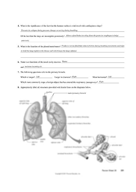
- Structure: It's reinforced with C-shaped cartilaginous rings, providing structural support while allowing flexibility during breathing. The inner lining is ciliated, helping to remove mucus and trapped particles.
B. The Bronchi and Bronchioles: The trachea branches into two main bronchi, one for each lung. These further subdivide into smaller and smaller bronchi, eventually leading to tiny bronchioles.
- Structure and Function: The bronchi, like the trachea, are supported by cartilage. As the bronchi branch into bronchioles, the cartilage gradually disappears, replaced by smooth muscle. This smooth muscle plays a vital role in regulating airflow through bronchoconstriction (narrowing) and bronchodilation (widening).
C. The Alveoli: The Sites of Gas Exchange: The terminal bronchioles lead to tiny air sacs called alveoli. These are the functional units of the respiratory system where gas exchange occurs.
- Structure and Function: Alveoli are surrounded by a network of capillaries, allowing for efficient diffusion of oxygen into the bloodstream and carbon dioxide out of the bloodstream. Their thin walls and large surface area maximize gas exchange.
D. The Lungs: The lungs are paired organs located in the thoracic cavity, protected by the rib cage and intercostal muscles. Each lung is divided into lobes: the right lung has three lobes, and the left lung has two.
- Structure and Function: The lungs are highly elastic, allowing them to expand and contract during breathing. They are covered by a serous membrane called the pleura, which helps to reduce friction during breathing. The pleura consists of two layers: the visceral pleura (covering the lungs) and the parietal pleura (lining the thoracic cavity). The space between these two layers, the pleural cavity, contains a small amount of fluid that lubricates the surfaces.
III. The Mechanics of Breathing: A Coordinated Effort
Breathing, or pulmonary ventilation, is the process of moving air into and out of the lungs. It involves several key muscles and structures:
A. Inspiration (Inhalation): This is an active process requiring muscle contraction.
- Diaphragm: The diaphragm, a dome-shaped muscle at the base of the thoracic cavity, contracts and flattens, increasing the volume of the chest cavity.
- External Intercostal Muscles: These muscles located between the ribs contract, pulling the ribs upward and outward, further increasing the chest cavity volume.
- Pressure Changes: The increase in chest cavity volume decreases the pressure inside the lungs, creating a pressure gradient that draws air into the lungs.
B. Expiration (Exhalation): This can be either an active or passive process.
- Passive Expiration: During quiet breathing, expiration is passive. The diaphragm and external intercostal muscles relax, causing the chest cavity to decrease in volume, increasing the pressure inside the lungs and forcing air out.
- Active Expiration: During forceful exhalation, such as during exercise or coughing, internal intercostal muscles and abdominal muscles contract, actively reducing the chest cavity volume and forcing air out.
IV. Control of Respiration: A Complex Regulatory System
Breathing is a finely tuned process regulated by the nervous and endocrine systems:
A. Nervous System Control: The respiratory center in the brainstem (medulla oblongata and pons) controls the rate and depth of breathing. This center receives input from chemoreceptors, which monitor blood levels of oxygen, carbon dioxide, and pH.
- Chemoreceptors: These sensors are located in the carotid bodies (in the neck) and the aortic bodies (in the aorta). They detect changes in blood gas levels and pH, sending signals to the respiratory center to adjust breathing accordingly. For example, an increase in carbon dioxide levels or a decrease in oxygen levels will trigger an increase in breathing rate and depth.
B. Endocrine System Influences: Hormones can also influence breathing, though to a lesser extent than the nervous system. For example, epinephrine (adrenaline) can increase breathing rate and depth in response to stress or exercise.
V. Clinical Considerations and Respiratory Health
Understanding the anatomy and physiology of the respiratory system is crucial for comprehending various respiratory conditions and promoting respiratory health.
A. Common Respiratory Disorders:
- Asthma: A chronic inflammatory disorder characterized by airway narrowing and bronchospasm.
- Chronic Obstructive Pulmonary Disease (COPD): A group of lung diseases including emphysema and chronic bronchitis, characterized by airflow limitation.
- Pneumonia: An infection of the lungs, often caused by bacteria, viruses, or fungi.
- Lung Cancer: A serious malignancy originating in the lungs.
B. Promoting Respiratory Health:
- Avoid Smoking: Smoking is a major risk factor for many respiratory diseases.
- Practice Good Hygiene: Frequent handwashing can help prevent respiratory infections.
- Get Vaccinated: Vaccination against influenza and pneumonia can help protect against these infections.
- Maintain a Healthy Lifestyle: Regular exercise, a balanced diet, and stress management can improve overall respiratory health.
VI. Conclusion:
The respiratory system is a complex and vital system that plays a crucial role in maintaining life. Understanding its anatomy, physiology, and the various factors that can affect its function is essential for promoting respiratory health and managing respiratory disorders. This detailed overview, expanding on the likely scope of “Exercise 36,” provides a solid foundation for further study and a deeper appreciation of this remarkable system. Remember that this information is for educational purposes and does not constitute medical advice. Always consult with a healthcare professional for any health concerns. Further exploration into specific diseases, diagnostic techniques, and treatment options will build upon this fundamental understanding of the respiratory system’s structure and function.
Latest Posts
Latest Posts
-
Translate The Medical Term Cerebral Thrombosis As Literally As Possible
Apr 08, 2025
-
An Introduction To Romanticism Mastery Test
Apr 08, 2025
-
How Can The Fishermen Save Gas
Apr 08, 2025
-
Tom Walker And The Devil Symbolism
Apr 08, 2025
-
Characters In In The Time Of The Butterflies
Apr 08, 2025
Related Post
Thank you for visiting our website which covers about Anatomy Of Respiratory System Exercise 36 . We hope the information provided has been useful to you. Feel free to contact us if you have any questions or need further assistance. See you next time and don't miss to bookmark.