Art-labeling Activity Anterior Muscles Of The Lower Body
Onlines
Mar 16, 2025 · 7 min read
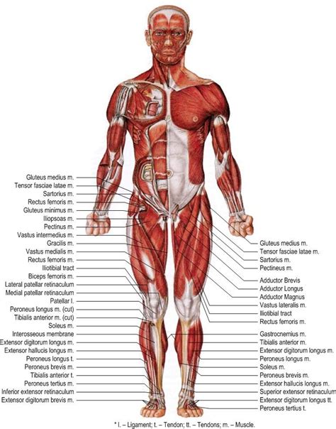
Table of Contents
Art-Labeling Activity: Anterior Muscles of the Lower Body
Understanding the anterior muscles of the lower body is crucial for anyone involved in anatomy, physical therapy, fitness training, or artistic representation of the human form. This comprehensive guide will delve into the intricacies of these muscles, exploring their functions, actions, and artistic representation through detailed labeling activities. We'll move beyond simple identification to a deeper understanding of muscle interaction and the nuanced ways they contribute to movement and overall form.
The Importance of Accurate Muscle Labeling
Accurate labeling of anatomical structures is paramount. In the artistic realm, it elevates the realism and credibility of anatomical drawings and sculptures. For those in healthcare professions, precise labeling is essential for effective communication, diagnosis, and treatment planning. This activity goes beyond simple memorization; it fosters a deeper understanding of muscle relationships, origins, insertions, and actions, enriching both artistic and scientific perspectives.
Anterior Lower Body Muscles: A Detailed Exploration
The anterior muscles of the lower body are primarily responsible for hip flexion, knee extension, and a range of other important movements. Let's explore each muscle group individually:
1. Iliopsoas Muscle Group
The iliopsoas is a powerful hip flexor, comprising two main muscles: the iliacus and the psoas major.
-
Iliacus: Originates from the iliac fossa of the hip bone and inserts into the lesser trochanter of the femur. Its primary action is hip flexion.
-
Psoas Major: Originates from the transverse processes of the lumbar vertebrae and inserts into the lesser trochanter of the femur. Besides hip flexion, it also contributes to lumbar spine flexion and lateral flexion.
Artistic Representation: When depicting the iliopsoas, focus on its deep position within the pelvis. It's not always visible superficially, but its powerful influence on hip movement needs to be acknowledged through the overall posture and position of the limb. Slight bulging in the groin region can suggest its tension.
Labeling Activity: Label the iliacus and psoas major on anatomical diagrams, paying close attention to their origins and insertions. Consider adding labels indicating their primary actions (hip flexion) and secondary actions (lumbar spine flexion for the psoas major).
2. Quadriceps Femoris Muscle Group
The quadriceps femoris is a group of four powerful muscles located on the anterior thigh, responsible primarily for knee extension.
-
Rectus Femoris: The only one of the quadriceps that crosses both the hip and knee joints. It originates from the anterior inferior iliac spine and the superior acetabulum of the hip bone, and inserts into the tibial tuberosity via the patellar tendon. Its actions include hip flexion and knee extension.
-
Vastus Lateralis: The largest of the quadriceps, originating from the greater trochanter, intertrochanteric line, and linea aspera of the femur. It inserts into the tibial tuberosity via the patellar tendon and its primary action is knee extension.
-
Vastus Medialis: Originates from the intertrochanteric line and medial linea aspera of the femur and inserts into the tibial tuberosity via the patellar tendon. Its primary action is knee extension.
-
Vastus Intermedius: Deep to the rectus femoris, originating from the anterior and lateral surfaces of the femur. It inserts into the tibial tuberosity via the patellar tendon. Its primary action is knee extension.
Artistic Representation: The quadriceps are prominent muscles that should be accurately represented in artworks. Their bulk and definition vary considerably depending on the individual's physique and level of muscle development. Consider the interplay of light and shadow to showcase the muscular contours and the distinct divisions between the four heads.
Labeling Activity: Label each of the four quadriceps muscles on anatomical drawings. Pay attention to their individual origins and insertions, highlighting the rectus femoris's dual function across both the hip and knee joints. Consider adding labels to show the patella and patellar tendon. Experiment with shading techniques to show the muscle's form and bulk.
3. Sartorius Muscle
The sartorius is a long, thin muscle that spans the entire length of the thigh.
- Origin: Anterior superior iliac spine
- Insertion: Medial surface of the proximal tibia (Pes Anserine)
- Actions: Hip flexion, abduction, and external rotation; knee flexion
Artistic Representation: The sartorius is a superficial muscle, making it relatively easy to represent. Its long, diagonal course across the thigh should be clearly visible. Its contribution to thigh movement, especially in situations involving crossing the legs or sitting with legs crossed, should be reflected in the artwork.
Labeling Activity: Label the sartorius muscle on anatomical diagrams, paying attention to its unique diagonal course. Include labels that indicate its multiple actions (hip flexion, abduction, and external rotation; knee flexion).
4. Pectineus Muscle
The pectineus is a small, flat muscle located medially on the thigh.
- Origin: Pectineal line of the pubis
- Insertion: Pectineal line of the femur
- Actions: Hip flexion, adduction, and medial rotation
Artistic Representation: While relatively small, the pectineus contributes to the overall form of the inner thigh. Its position should be accurately reflected, especially in poses that involve adduction of the hip. Subtle changes in the inner thigh’s contour can suggest its role.
Labeling Activity: Label the pectineus on anatomical diagrams. Consider including labels highlighting its role in hip adduction and medial rotation, especially when comparing it to the other hip adductors.
5. Gracilis Muscle
The gracilis is a long, thin muscle located on the medial side of the thigh.
- Origin: Inferior pubic ramus
- Insertion: Medial surface of the proximal tibia (Pes Anserine)
- Actions: Hip adduction, flexion, and medial rotation; knee flexion
Artistic Representation: Like the sartorius, the gracilis's long, slender form is relatively straightforward to represent. Its position along the medial thigh should be clear. It's important to show its relationship with the sartorius and the other muscles inserting at the Pes Anserine.
Labeling Activity: Label the gracilis muscle, noting its location on the medial thigh and its insertion at the Pes Anserine, alongside the sartorius and semitendinosus. Highlight its actions, particularly hip adduction and knee flexion.
Enhancing Artistic Representation through Understanding Muscle Interactions
The true mastery of anatomical art lies not just in labeling individual muscles but in understanding their synergistic and antagonistic relationships. For example, the quadriceps (knee extensors) and hamstrings (knee flexors) work antagonistically; when one contracts, the other relaxes. Depicting this dynamic relationship realistically adds layers of depth and realism to your artwork.
Understanding how muscles work together to create movement is key. Consider the interplay of the iliopsoas, sartorius, and quadriceps during a walking motion. Or visualize the coordinated actions of the pectineus, gracilis, and adductor muscles during a lateral leg movement. Observing real-life movements and breaking them down into the contributing muscle groups can significantly improve your artistic representation.
Advanced Labeling Activities: Beyond Basic Identification
To further enhance your understanding and artistic skill, consider these advanced labeling activities:
- Muscle Layer Drawings: Create layered drawings, starting with the deepest muscles and progressively adding superficial layers. This helps visualize the spatial relationships between muscles.
- Movement-Based Drawings: Create drawings depicting specific actions, such as a high kick or a deep squat. Label the muscles actively involved in generating those movements.
- Comparative Anatomy: Compare the anterior lower body musculature of different species. This broader perspective enhances understanding of functional adaptations and variations.
- Dynamic Contraction Drawings: Depict muscles in various stages of contraction. Show the changes in muscle shape and volume during different phases of movement. This requires a deeper understanding of muscle physiology.
- Artistic Exploration: Explore different artistic mediums to represent the muscles. Experiment with pencil sketches, charcoal drawings, paintings, or even three-dimensional sculptures to better visualize and appreciate their form.
Conclusion: The Synergistic Relationship between Art and Anatomy
Art-labeling activities, especially focusing on complex anatomical structures like the anterior muscles of the lower body, provide a powerful tool for both artistic and scientific understanding. By combining precise labeling with an appreciation for muscle interactions and dynamic movement, artists and scientists alike can significantly enhance their knowledge and artistic representation of the human form. This detailed exploration should serve as a springboard for ongoing learning and creative expression. The more you delve into the intricacies of the human anatomy, the more nuanced and compelling your artistic representations will become. Continuous exploration and practical application are crucial to mastering this fascinating field.
Latest Posts
Latest Posts
-
Tuesdays With Morrie Summary Per Chapter
Mar 17, 2025
-
Esta Manana Comi Frutas En El
Mar 17, 2025
-
10 2 3 Select And Configure Dual Monitors
Mar 17, 2025
-
The Dutch Hunger Winter Case Study Answers
Mar 17, 2025
-
Journey To The Center Of The Earth Book Notes
Mar 17, 2025
Related Post
Thank you for visiting our website which covers about Art-labeling Activity Anterior Muscles Of The Lower Body . We hope the information provided has been useful to you. Feel free to contact us if you have any questions or need further assistance. See you next time and don't miss to bookmark.
