Art-labeling Activity Anterior Muscles Of The Upper Body
Onlines
Apr 06, 2025 · 6 min read
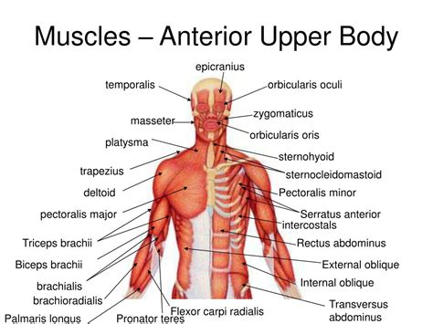
Table of Contents
Art-Labeling Activity: Anterior Muscles of the Upper Body
Art-labeling activities offer a dynamic and engaging approach to learning human anatomy, particularly the intricate network of muscles. This article delves into the anterior muscles of the upper body, providing a detailed description suitable for both students and enthusiasts. We'll explore each muscle's origin, insertion, action, and innervation, enhanced by visual aids and practical labeling exercises. This approach strengthens understanding through active learning and visual reinforcement, vital for effective memorization and application.
Understanding the Anterior Muscles: A Functional Overview
The anterior muscles of the upper body primarily govern movements related to flexion, adduction, and medial rotation of the shoulder and elbow joints. They also play crucial roles in respiration and posture. This group includes muscles spanning the chest, shoulder, and arm, each contributing uniquely to the upper body's complex movement repertoire. Their synergistic and antagonistic interactions enable a vast range of precise and powerful actions.
Key Muscle Groups and Their Actions:
-
Pectoralis Major: This large, fan-shaped muscle dominates the chest. It's responsible for adduction, flexion, and medial rotation of the humerus (upper arm bone). It also assists in forced inspiration.
-
Pectoralis Minor: Located beneath the pectoralis major, this muscle draws the scapula (shoulder blade) forward and downward, contributing to shoulder girdle movements.
-
Serratus Anterior: Situated on the lateral chest wall, this muscle protracts the scapula, moving it away from the spine. It also plays a critical role in upward rotation of the scapula.
-
Subclavius: A small muscle located beneath the clavicle (collarbone), it stabilizes and depresses the clavicle.
-
Biceps Brachii: The familiar "bicep" muscle, located on the anterior arm, flexes the elbow and supinates the forearm. It also weakly assists in shoulder flexion.
-
Brachialis: Deep to the biceps brachii, this muscle is the primary flexor of the elbow.
-
Brachioradialis: Located on the lateral side of the forearm, it flexes the elbow, especially when the forearm is in a neutral position.
-
Coracobrachialis: A small muscle originating from the coracoid process of the scapula, it flexes and adducts the shoulder.
Art-Labeling Exercises: Enhancing Learning Through Visualization
The most effective way to learn the anterior muscles of the upper body is through active engagement. Art labeling is an excellent method combining visual learning with practical application. Here are several exercises to improve your understanding:
Exercise 1: Basic Muscle Identification
Materials: A high-quality anatomical illustration or diagram of the anterior muscles of the upper body.
Instructions:
- Print: Print a clear image, ensuring it’s large enough for comfortable labeling.
- Label: Using a pen or pencil, carefully label each muscle listed above on the diagram. Pay close attention to the precise location of each muscle’s origin and insertion.
- Verification: Check your work against a reliable anatomical text or online resource. Correct any mistakes.
Exercise 2: Muscle Origin and Insertion
Materials: The same anatomical illustration used in Exercise 1.
Instructions:
- Identify Origins and Insertions: For each muscle, identify and label its origin (where it starts) and insertion (where it attaches). This will require a deeper understanding of skeletal anatomy.
- Color-Coding: Use different colored pens or pencils to highlight origins and insertions for each muscle. This will aid in visualization of muscle movement.
- Detailed Notes: Write brief notes next to each labeled muscle, summarizing its origin, insertion, and primary action.
Exercise 3: Muscle Action and Innervation
Materials: The anatomical illustration and a table summarizing the innervation of each muscle (nerves supplying the muscle).
Instructions:
- Action Description: For each muscle, write a brief description of its primary actions. Include any secondary actions or synergistic relationships with other muscles.
- Innervation: Identify and label the nerve that innervates each muscle. This will enhance your understanding of the neurological control of movement.
- Movement Scenarios: Imagine performing different upper body movements (e.g., pushing a heavy object, throwing a ball). Identify which muscles are primarily involved in each movement.
Exercise 4: Advanced Labeling: Muscle Layers and Relationships
Materials: Anatomical illustrations showing different layers of anterior muscles.
Instructions:
- Layer Identification: Identify and label superficial and deep muscle layers.
- Spatial Relationships: Describe the spatial relationships between muscles; which muscles lie superficial to others? Which muscles overlap?
- Functional Implications: Explain how the layering of muscles contributes to their coordinated function.
Exercise 5: Creating Your Own Anatomical Illustration
Materials: Blank paper or a digital drawing program.
Instructions:
- Sketch: Attempt to draw the anterior muscles of the upper body from memory, focusing on their relative size, shape, and location.
- Labeling: Label each muscle you’ve drawn, including its origin, insertion, and primary action.
- Refinement: Compare your drawing to a reference image and refine your work based on any discrepancies. This exercise significantly improves memorization through active recall.
Advanced Considerations: Clinical Applications and Further Learning
Understanding the anterior muscles of the upper body extends beyond anatomical knowledge; it holds critical importance in various clinical settings. Injuries to these muscles are common, particularly in athletes and individuals performing physically demanding tasks. Diagnosing and treating such injuries require a thorough grasp of muscle anatomy, function, and biomechanics.
Clinical Relevance:
- Shoulder impingement: Understanding the relationships between the rotator cuff muscles, pectoralis muscles, and the subacromial space is crucial for diagnosing and managing shoulder impingement syndrome.
- Rotator cuff tears: Knowledge of the actions and interactions of the anterior shoulder muscles helps in rehabilitation strategies following rotator cuff injuries.
- Biceps tendonitis: Accurate diagnosis and treatment of biceps tendonitis require detailed knowledge of the biceps brachii’s attachments and function.
- Carpal tunnel syndrome: While not directly related to the anterior upper body muscles, the understanding of forearm muscle anatomy helps identify potential contributing factors.
Further Learning:
- Palpation: Practicing palpation (feeling the muscles) on yourself or a partner enhances your understanding of muscle location and shape.
- Clinical Anatomy Texts: Supplement your learning with detailed clinical anatomy textbooks that provide deeper insights into muscle function and clinical relevance.
- Online Resources: Numerous reputable online resources, including anatomical atlases and interactive tutorials, offer further learning opportunities.
Conclusion: Mastering the Anterior Muscles Through Active Engagement
Art-labeling activities provide a powerful learning tool for mastering the intricacies of the anterior muscles of the upper body. Through active engagement and visual reinforcement, these exercises transform passive learning into a dynamic and memorable experience. The application of this knowledge extends far beyond academic understanding, proving invaluable in clinical settings and various physical activity applications. Consistent practice and a dedicated approach will lead to a robust understanding of this crucial anatomical region. Remember to always consult reliable anatomical resources and consider seeking guidance from experienced professionals when applying this knowledge in clinical or practical settings.
Latest Posts
Latest Posts
-
Lab 1 3 Testing Mode Identify Internal Components Of A Computer
Apr 07, 2025
-
What Is The Theme Of Julius Caesar
Apr 07, 2025
-
Health Chapter 22 Review Answer Key
Apr 07, 2025
-
You Are Transporting A Load Of Hazmat Across The Plains
Apr 07, 2025
-
Add A New Calculated Field Named Tuition
Apr 07, 2025
Related Post
Thank you for visiting our website which covers about Art-labeling Activity Anterior Muscles Of The Upper Body . We hope the information provided has been useful to you. Feel free to contact us if you have any questions or need further assistance. See you next time and don't miss to bookmark.
