Art-labeling Activity: Overview Of The Cardiac Conduction System
Onlines
Mar 24, 2025 · 7 min read
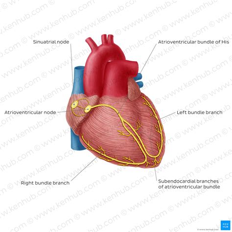
Table of Contents
Art-Labeling Activity: An Overview of the Cardiac Conduction System
The human heart, a tireless muscle, beats rhythmically throughout our lives, pumping blood to every corner of our body. This remarkable feat is orchestrated by a specialized network of cells known as the cardiac conduction system. Understanding this intricate system is crucial for comprehending normal heart function and diagnosing a wide array of cardiac pathologies. This article will serve as a comprehensive guide to the cardiac conduction system, utilizing an art-labeling activity approach to enhance learning and retention. We will explore each component, its role, and the potential consequences of dysfunction.
The Pacemaker of the Heart: The Sinoatrial (SA) Node
The journey begins with the sinoatrial (SA) node, often referred to as the heart's natural pacemaker. Located in the right atrium, near the superior vena cava, the SA node is a cluster of specialized cells capable of spontaneously generating electrical impulses. These impulses are rhythmic and initiate the heartbeat, setting the pace for the entire cardiac cycle. The SA node's inherent rhythmicity is determined by the balance of ion channels and their associated currents, leading to spontaneous depolarization and repolarization cycles.
Activity: Imagine drawing a simple heart. Label the location of the SA node with a star and write "SA Node - Heart's Pacemaker" next to it.
SA Node Dysfunction: Bradycardia and Tachycardia
Proper functioning of the SA node is critical. If the SA node fails to generate impulses at the normal rate, it can lead to bradycardia, a slow heart rate. Conversely, if the SA node generates impulses too rapidly, it results in tachycardia, a fast heart rate. Both bradycardia and tachycardia can be symptomatic and indicative of underlying cardiac issues.
The Atrioventricular (AV) Node: Gateway to the Ventricles
After the electrical impulse originates in the SA node, it travels through the atrial myocardium, causing atrial contraction. The impulse then reaches the atrioventricular (AV) node, situated in the right atrium near the tricuspid valve. The AV node plays a crucial role as the gatekeeper between the atria and the ventricles. It delays the impulse briefly, allowing the atria to fully contract and empty their blood into the ventricles before ventricular contraction begins. This delay ensures efficient blood flow.
Activity: On your heart drawing, label the AV node with a circle and write "AV Node - Gateway to Ventricles". Draw an arrow indicating the pathway of the impulse from the SA node to the AV node.
AV Node Block: Disruption of Conduction
Atrioventricular (AV) block occurs when the conduction of the electrical impulse through the AV node is impaired or interrupted. This can result in varying degrees of heart block, from mild first-degree AV block to complete heart block, where no impulses are conducted from the atria to the ventricles. AV blocks necessitate careful evaluation and management.
The Bundle of His: The Only Electrical Connection Between Atria and Ventricles
Following the AV node, the electrical impulse travels down the bundle of His, also known as the atrioventricular bundle. This is the only electrical connection between the atria and the ventricles. The bundle of His is a specialized conducting pathway that rapidly transmits the impulse from the AV node to the ventricles. Its location within the interventricular septum ensures efficient conduction to both ventricles.
Activity: Add the Bundle of His to your drawing, labeling it with a thick line connecting the AV node to the Bundle Branches. Note its location within the interventricular septum.
Bundle Branch Block: Impaired Ventricular Conduction
Bundle branch blocks occur when conduction through one of the bundle branches is impaired or blocked. This can lead to asynchrony in ventricular contraction, affecting the efficiency of the heart's pumping action. Left bundle branch block (LBBB) and right bundle branch block (RBBB) are common types, often associated with underlying cardiac disease.
Bundle Branches: Ensuring Coordinated Ventricular Contraction
The bundle of His divides into two major branches: the right bundle branch and the left bundle branch. These branches further divide into smaller Purkinje fibers, ensuring the impulse reaches all parts of the ventricles simultaneously. This coordinated conduction is essential for synchronized ventricular contraction and efficient ejection of blood.
Activity: On your drawing, show the branching of the Bundle of His into the right and left bundle branches, extending towards the apex of the heart. Label each branch.
Bundle Branch Blocks and Their Clinical Significance
As previously mentioned, bundle branch blocks can significantly impact ventricular depolarization. This can lead to a variety of symptoms, including palpitations, shortness of breath, and even syncope (fainting). Electrocardiography (ECG) is instrumental in diagnosing these blocks.
Purkinje Fibers: Rapid Conduction Throughout the Ventricles
The final component of the conduction system are the Purkinje fibers. These fine, branching fibers spread throughout the ventricular myocardium, ensuring rapid and efficient distribution of the electrical impulse. This allows for near-simultaneous contraction of the ventricles, maximizing the ejection of blood into the pulmonary artery and aorta. The Purkinje fibers possess a high density of gap junctions, facilitating rapid conduction.
Activity: Add numerous smaller branches extending from the bundle branches to represent the Purkinje fibers. Label them accordingly.
Purkinje Fiber Dysfunction: Ventricular Arrhythmias
Disruptions in Purkinje fiber function can lead to various ventricular arrhythmias, including premature ventricular contractions (PVCs) and potentially life-threatening ventricular tachycardia and fibrillation. These arrhythmias can disrupt the heart's normal rhythm and compromise its pumping ability.
The Interplay and Importance of Each Component
The cardiac conduction system works as a coordinated unit. Each component plays a critical role in generating and transmitting the electrical impulse that drives the heartbeat. A disruption in any part of this system can have significant consequences, ranging from mild symptoms to life-threatening arrhythmias. Therefore, understanding the function and interdependence of each component is crucial for clinicians and healthcare professionals.
Activity: Review your drawing. Trace the pathway of the electrical impulse from the SA node to the Purkinje fibers, highlighting the sequential activation of each component. Consider adding arrows to illustrate the direction of impulse conduction.
Clinical Significance and Diagnostic Tools
Numerous diagnostic tools are used to assess the cardiac conduction system. Electrocardiography (ECG) is a fundamental test that provides a real-time representation of the heart's electrical activity. ECG can identify various conduction abnormalities, including bradycardia, tachycardia, heart blocks, and bundle branch blocks. Echocardiography provides anatomical information about the heart and can help identify structural abnormalities that might affect conduction. Electrophysiological studies (EPS) are more invasive procedures involving the insertion of catheters to directly study the heart's electrical activity. EPS can identify specific locations of conduction abnormalities and guide therapeutic interventions.
Therapeutic Interventions for Conduction System Disorders
Treatment for cardiac conduction system disorders depends on the specific condition and its severity. Pacemakers are implantable devices that provide electrical stimulation to the heart, correcting slow heart rates (bradycardia). Implantable cardioverter-defibrillators (ICDs) are used to detect and treat life-threatening arrhythmias such as ventricular tachycardia and fibrillation. Medication can be used to manage various conduction disorders, modifying heart rate and rhythm. In some cases, surgical interventions may be necessary to address underlying structural abnormalities that affect conduction.
Conclusion: The Art of Understanding the Heart
The cardiac conduction system is a fascinating and complex network that governs the rhythmic beating of our heart. By utilizing an art-labeling approach, we have explored the anatomy and physiology of this essential system, highlighting the critical roles of each component and the clinical significance of its dysfunction. This detailed overview not only enhances comprehension but also underscores the importance of continued research and advancement in the diagnosis and management of cardiac conduction disorders. This knowledge empowers us to better appreciate the intricate mechanisms that maintain our cardiovascular health. Remember, regular check-ups, a healthy lifestyle, and timely medical attention are crucial for preserving the well-being of our remarkable hearts.
Latest Posts
Latest Posts
-
Windows Picture Password Belongs To Which Of The Following
Mar 26, 2025
-
For Each Graph Select All Symmetries That Apply
Mar 26, 2025
-
Theme Of All Summer In A Day
Mar 26, 2025
-
The Joy Luck Club Chapter 1 Summary
Mar 26, 2025
-
Encyclopedia Pages Should Never Get Low Needs Met Ratings
Mar 26, 2025
Related Post
Thank you for visiting our website which covers about Art-labeling Activity: Overview Of The Cardiac Conduction System . We hope the information provided has been useful to you. Feel free to contact us if you have any questions or need further assistance. See you next time and don't miss to bookmark.
