Cell Division Worksheet #1 Microscope Images
Onlines
Mar 24, 2025 · 6 min read
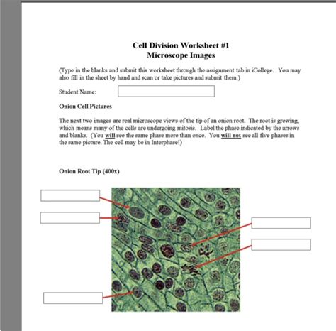
Table of Contents
Cell Division Worksheet #1: Microscope Images - A Deep Dive into Mitosis and Meiosis
Understanding cell division, particularly mitosis and meiosis, is fundamental to grasping the intricacies of biology. This worksheet utilizes microscope images to guide you through the key stages of these crucial cellular processes. We will delve into the details of each phase, highlighting the distinguishing features and providing a framework for accurate identification. By the end of this comprehensive guide, you will be proficient in analyzing microscopic images of cell division, understanding the significance of each phase, and appreciating the underlying mechanisms that drive the continuity of life.
Understanding the Basics: Mitosis and Meiosis
Before we jump into analyzing the microscope images, let's refresh our understanding of mitosis and meiosis. Both are types of cell division, but they serve vastly different purposes.
Mitosis: The Foundation of Growth and Repair
Mitosis is a type of cell division that results in two daughter cells, each having the same number and kind of chromosomes as the parent cell. This process is essential for:
- Growth: Mitosis allows multicellular organisms to grow from a single fertilized egg to a complex organism with trillions of cells.
- Repair: It replaces damaged or worn-out cells, ensuring the integrity of tissues and organs.
- Asexual Reproduction: In some organisms, mitosis is the primary method of asexual reproduction, creating genetically identical offspring.
Meiosis: The Basis of Sexual Reproduction
Meiosis is a specialized type of cell division that reduces the chromosome number by half, producing four haploid daughter cells (gametes). This process is crucial for:
- Sexual Reproduction: Meiosis generates the genetic diversity necessary for sexual reproduction by creating gametes (sperm and egg cells) with unique combinations of chromosomes.
- Genetic Variation: The shuffling and recombination of genetic material during meiosis contribute to the incredible diversity observed within populations.
Analyzing Microscope Images: A Step-by-Step Guide
Analyzing microscope images of cell division requires careful observation and a systematic approach. The following steps will guide you through the process:
1. Identify the Type of Cell Division
The first step is determining whether the images depict mitosis or meiosis. Mitosis typically shows a single cell undergoing division, resulting in two identical daughter cells. Meiosis, on the other hand, involves two sequential divisions (Meiosis I and Meiosis II) and results in four genetically diverse daughter cells. Look for clues such as the number of chromosomes and the presence of homologous pairs.
2. Identify the Stages of Cell Division
Once you've identified the type of cell division, focus on identifying the specific stages. Below is a breakdown of the key stages, including features to look for in microscope images:
Mitosis:
- Prophase: Chromosomes condense and become visible, the nuclear envelope breaks down, and the mitotic spindle begins to form. Look for: Thick, condensed chromosomes; disappearance of the nuclear membrane.
- Metaphase: Chromosomes align along the metaphase plate (the equator of the cell). Look for: Chromosomes arranged in a single line across the middle of the cell.
- Anaphase: Sister chromatids separate and move to opposite poles of the cell. Look for: V-shaped chromosomes moving towards opposite ends of the cell.
- Telophase: Chromosomes decondense, the nuclear envelope reforms, and the cytoplasm begins to divide (cytokinesis). Look for: Two distinct nuclei forming; chromosomes becoming less visible.
- Cytokinesis: The final stage where the cytoplasm divides, resulting in two separate daughter cells. Look for: A cleavage furrow (in animal cells) or a cell plate (in plant cells) forming.
Meiosis I:
- Prophase I: Homologous chromosomes pair up (synapsis) and crossing over occurs, exchanging genetic material. Look for: Paired homologous chromosomes; chiasmata (points of crossing over).
- Metaphase I: Homologous chromosome pairs align along the metaphase plate. Look for: Paired chromosomes arranged in a line, unlike the individual chromosomes in mitosis metaphase.
- Anaphase I: Homologous chromosomes separate and move to opposite poles. Look for: Movement of entire chromosomes, not sister chromatids, to opposite poles.
- Telophase I: The cytoplasm divides (cytokinesis), resulting in two haploid daughter cells. Look for: Two cells with half the number of chromosomes as the original cell.
Meiosis II:
Meiosis II is very similar to mitosis, except that it starts with haploid cells. The stages are:
- Prophase II: Chromosomes condense.
- Metaphase II: Chromosomes align along the metaphase plate.
- Anaphase II: Sister chromatids separate and move to opposite poles.
- Telophase II: The cytoplasm divides (cytokinesis), resulting in four haploid daughter cells.
3. Annotate and Label Your Images
Once you've identified the stages, annotate your microscope images, labeling the key structures such as chromosomes, spindle fibers, centrioles (if visible), and the nuclear envelope. This will reinforce your understanding and provide a clear record of your analysis.
Practical Application: Interpreting Sample Microscope Images
Let's now analyze some hypothetical microscope images, applying the knowledge we’ve gained. Remember, these are examples, and real images may vary slightly depending on the organism and staining techniques used.
Image 1: Shows condensed chromosomes aligned along the center of the cell. Sister chromatids are clearly visible, attached at the centromere. No homologous pairs are evident.
Analysis: This image depicts mitosis, specifically metaphase. The alignment of individual chromosomes at the metaphase plate indicates this stage. The absence of homologous pairs rules out meiosis.
Image 2: Shows two cells, each with half the number of chromosomes compared to the original cell. Chromosomes are less condensed than in the previous image.
Analysis: This image depicts telophase I of meiosis. The formation of two cells, each with a haploid chromosome number, is characteristic of the end of Meiosis I.
Image 3: Shows paired homologous chromosomes, with visible chiasmata (crossing over points). The nuclear envelope is dissolving.
Analysis: This image depicts prophase I of meiosis. The presence of homologous chromosome pairs undergoing synapsis and crossing over is a key feature of prophase I.
Advanced Considerations
- Chromosome Number: Pay close attention to the chromosome number in the images. This is a crucial factor in distinguishing between mitosis and meiosis, as well as determining the species of organism being studied.
- Species Variation: The appearance of chromosomes and the details of cell division can vary slightly depending on the species of organism.
- Staining Techniques: Different staining techniques will affect the visibility of various cellular structures.
Conclusion
Analyzing microscope images of cell division is a powerful way to learn about the intricacies of mitosis and meiosis. By following a systematic approach and paying close attention to the key features of each stage, you can accurately identify the stages of cell division and gain a deeper understanding of these fundamental biological processes. Remember to practice regularly and consult reliable resources to enhance your skill in interpreting these important images. This deep understanding of cell division is vital for future study in genetics, developmental biology, and cancer research. Continuous practice and review will further solidify your ability to confidently and accurately analyze microscope images of cell division, paving the way for a stronger grasp of the fundamentals of biology. The skills learned here are applicable to a vast array of biological studies, showcasing the importance of this topic and its practical implications.
Latest Posts
Latest Posts
-
The Clinical Protocol Must Identify Alternatives To Restraints
Mar 30, 2025
-
Conversion Factors And Problem Solving Lab 2 Report Sheet
Mar 30, 2025
-
Anti Vietnam War Movement Graphic Organizer
Mar 30, 2025
-
Discuss The Importance Of Legislation To Protect Marshes
Mar 30, 2025
-
Symbols In All The Light We Cannot See
Mar 30, 2025
Related Post
Thank you for visiting our website which covers about Cell Division Worksheet #1 Microscope Images . We hope the information provided has been useful to you. Feel free to contact us if you have any questions or need further assistance. See you next time and don't miss to bookmark.
