Color A Typical Prokaryote Cell Answer Key
Onlines
Mar 20, 2025 · 6 min read
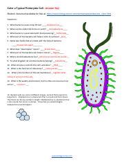
Table of Contents
Coloring a Typical Prokaryote Cell: A Detailed Guide with Answer Key
Understanding prokaryotic cell structure is fundamental to grasping the basics of biology. This comprehensive guide provides a detailed walkthrough of coloring a typical prokaryote cell, complete with an answer key and insightful explanations to reinforce your learning. We will delve into the intricacies of prokaryotic cell anatomy, focusing on the key structures you'll be identifying and coloring. Mastering this exercise will solidify your understanding of these essential life forms.
Understanding Prokaryotic Cells: A Quick Overview
Before we dive into the coloring exercise, let's refresh our understanding of prokaryotic cells. Prokaryotes are single-celled organisms that lack a membrane-bound nucleus and other membrane-bound organelles. This distinguishes them from eukaryotes, which possess a nucleus and other complex organelles. Prokaryotes are incredibly diverse, including bacteria and archaea, and play crucial roles in various ecosystems.
Key Features of Prokaryotic Cells You'll Be Coloring:
- Plasma Membrane (Cell Membrane): This outermost boundary regulates the passage of substances into and out of the cell. Think of it as the cell's gatekeeper.
- Cytoplasm: The jelly-like substance filling the cell, containing the cell's components. It's the bustling hub of cellular activity.
- Nucleoid: The region where the prokaryotic DNA is located. Although not membrane-bound like a eukaryotic nucleus, it's still crucial for genetic information storage.
- Ribosomes: The protein synthesis machinery of the cell. These are responsible for creating proteins, essential for various cellular functions.
- Cell Wall: A rigid outer layer that provides structural support and protection to the cell. The composition of the cell wall differs between bacteria and archaea.
- Capsule (Optional): Some prokaryotes have a capsule, a sticky outer layer that contributes to adhesion and protection. This is not always present.
- Pili (Optional): Hair-like appendages used for attachment to surfaces or for conjugation (exchange of genetic material). These are not always present.
- Flagella (Optional): Whip-like structures used for motility – the ability to move around. Not all prokaryotes possess flagella.
The Coloring Exercise: A Step-by-Step Guide
Now, let's move onto the coloring exercise itself. For the purpose of this guide, we'll use a simplified representation of a typical prokaryotic cell. You can adapt this to more complex diagrams as your understanding grows.
Materials You'll Need:
- A printed diagram of a prokaryotic cell (you can easily find these online or create your own based on the descriptions above).
- Colored pencils, crayons, or markers.
Coloring Instructions:
-
Plasma Membrane (Cell Membrane): Color this structure a vibrant blue. This will help visually distinguish the cell's boundary.
-
Cytoplasm: Fill the interior of the cell with a light yellow or pale green. This represents the cellular fluid within which all other components reside.
-
Nucleoid: Color the nucleoid region a darker shade of green. This highlights the area where the prokaryotic DNA is concentrated.
-
Ribosomes: Use red dots or small circles to represent ribosomes scattered throughout the cytoplasm. Their small size emphasizes their abundance within the cell.
-
Cell Wall: Color the cell wall a solid brown. This emphasizes its rigid nature and protective function.
-
Capsule (if present): If your diagram includes a capsule, color it a light pink. This represents the outermost layer of protection.
-
Pili (if present): If pili are present, color them purple. This will help distinguish these attachment structures.
-
Flagella (if present): If flagella are present, color them orange. This highlights their role in movement.
Answer Key and Detailed Explanations:
Below, we provide a detailed explanation for each structure, reinforcing its function and importance. You can compare your coloring with this answer key to ensure accuracy.
| Structure | Color | Function and Explanation |
|---|---|---|
| Plasma Membrane (Cell Membrane) | Blue | This selectively permeable barrier controls the movement of substances in and out of the cell. It's crucial for maintaining cellular homeostasis. |
| Cytoplasm | Pale Yellow/Green | The cytoplasm is the site of many metabolic reactions. It suspends the cell's organelles and provides a medium for cellular processes. |
| Nucleoid | Dark Green | This region contains the cell's genetic material (DNA), which dictates the cell's activities and characteristics. Note: it is not membrane-bound. |
| Ribosomes | Red Dots | Ribosomes are responsible for protein synthesis – creating proteins essential for structure and function. They are found throughout the cytoplasm. |
| Cell Wall | Brown | The rigid cell wall provides structural support and protection to the cell, preventing osmotic lysis (bursting) in hypotonic environments. Its composition varies between bacteria and archaea. |
| Capsule (if present) | Light Pink | The capsule is an outer layer made of polysaccharides or glycoproteins. It enhances adhesion to surfaces and protects against phagocytosis (engulfment by immune cells). |
| Pili (if present) | Purple | Pili are short, hair-like appendages used for attachment to surfaces or for bacterial conjugation (transfer of genetic material). |
| Flagella (if present) | Orange | Flagella are long, whip-like structures that enable the cell to move through its environment. Their movement is driven by the rotation of basal bodies. |
Advanced Considerations: Beyond the Basics
While this exercise focuses on a simplified model, it's essential to appreciate the diversity within prokaryotes. Variations exist in cell wall composition (Gram-positive vs. Gram-negative bacteria), presence of additional structures (e.g., plasmids, inclusion bodies), and overall cell morphology (shape – coccus, bacillus, spirillum).
Gram-Positive vs. Gram-Negative Bacteria: A Deeper Dive
The Gram stain is a crucial technique in microbiology that distinguishes between two major types of bacteria based on their cell wall structure.
-
Gram-positive bacteria: These possess a thick peptidoglycan layer in their cell wall, which retains the crystal violet dye in the Gram staining procedure. This results in a purple coloration.
-
Gram-negative bacteria: These have a thinner peptidoglycan layer and an outer membrane containing lipopolysaccharide (LPS). The crystal violet dye is easily washed away, revealing a pink coloration after counterstaining with safranin.
This difference in cell wall structure is significant, impacting their susceptibility to antibiotics and their interactions with the immune system.
Exploring Additional Prokaryotic Structures:
Beyond the structures covered in the basic coloring exercise, several other features might be found in prokaryotic cells:
-
Plasmids: Small, circular DNA molecules that replicate independently of the chromosomal DNA. They often carry genes conferring antibiotic resistance or other advantageous traits.
-
Inclusion Bodies: Storage granules of various substances, such as glycogen (energy storage), polyphosphate (phosphate storage), or sulfur granules.
-
Endospores: Highly resistant dormant structures formed by some bacteria under stressful conditions. They can survive extreme temperatures, desiccation, and radiation.
Understanding these additional structures enhances your comprehension of the complexity and adaptability of prokaryotic cells.
Conclusion: Mastering Prokaryotic Cell Structure
Coloring a prokaryotic cell is a valuable exercise that helps solidify your understanding of its fundamental components and their functions. By carefully identifying and coloring each structure, you not only enhance your visual learning but also develop a deeper appreciation for the complexity and diversity within these microscopic organisms. Remember that this exercise serves as a foundational step—there's a vast world of prokaryotic biology waiting to be explored! Continue your learning by delving into the intricacies of prokaryotic metabolism, genetics, and their ecological significance. By building upon this foundation, you'll be well-equipped to tackle more advanced concepts in microbiology and related fields.
Latest Posts
Latest Posts
-
Discovery Channel Body Story Breaking Down Answer Key
Mar 21, 2025
-
Rn Learning System Communication Final Quiz
Mar 21, 2025
-
Sedra Smith Microelectronic Circuits 8th Edition Solutions Pdf
Mar 21, 2025
-
Interactive Tutorial Forming Questions In Spanish
Mar 21, 2025
-
Tienes Tu Cuaderno No No 1 Of 1 Tengo
Mar 21, 2025
Related Post
Thank you for visiting our website which covers about Color A Typical Prokaryote Cell Answer Key . We hope the information provided has been useful to you. Feel free to contact us if you have any questions or need further assistance. See you next time and don't miss to bookmark.
