Concept Map Body Cavities And Membranes
Onlines
Mar 07, 2025 · 6 min read
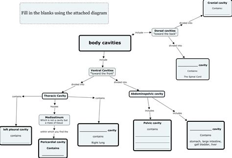
Table of Contents
Concept Map: Body Cavities and Membranes
Understanding the organization of the human body is crucial for comprehending anatomy and physiology. A key aspect of this organization involves the body cavities and the membranes that line them. This article provides a comprehensive overview of this topic, utilizing a concept map approach to illustrate the relationships between different structures and their functions. We will explore the major body cavities, their subdivisions, the serous membranes, and their clinical significance.
Major Body Cavities: A Hierarchical Overview
The human body is broadly divided into two major cavities: the dorsal cavity and the ventral cavity. These cavities house and protect vital organs.
1. The Dorsal Cavity: Protecting the Central Nervous System
The dorsal cavity is located on the posterior side of the body and is further subdivided into two smaller cavities:
-
Cranial Cavity: This cavity is located within the skull and houses the brain, a critical organ responsible for controlling virtually all body functions. The brain's delicate structure is protected by the skull bones and the cerebrospinal fluid. Meninges, a three-layered membrane (dura mater, arachnoid mater, and pia mater), provide additional protection and support.
-
Vertebral (Spinal) Cavity: Extending from the cranial cavity down the vertebral column lies the vertebral cavity. This cavity houses the spinal cord, a crucial component of the central nervous system responsible for transmitting signals between the brain and the rest of the body. The spinal cord is also protected by the vertebrae and the meninges. Injury to the vertebrae can compromise the integrity of the spinal cord, leading to serious neurological deficits.
2. The Ventral Cavity: Housing Viscera and Enabling Movement
The ventral cavity, situated on the anterior side of the body, is much larger than the dorsal cavity and houses most of the internal organs, collectively known as viscera. It is further subdivided into the thoracic cavity and the abdominopelvic cavity.
-
Thoracic Cavity: Located superior to the diaphragm, the thoracic cavity contains the heart, lungs, and major blood vessels. It is further subdivided into three smaller cavities:
-
Pleural Cavities (2): Each lung resides within its own pleural cavity, lined by a serous membrane called the pleura. The pleura consists of two layers: the visceral pleura adheres to the lung surface, and the parietal pleura lines the thoracic wall. The space between these layers is the pleural space, which contains a small amount of serous fluid that lubricates the surfaces, reducing friction during breathing. Pleurisy, an inflammation of the pleura, can cause significant pain and impair breathing.
-
Pericardial Cavity: The heart is enclosed within the pericardial cavity, surrounded by a serous membrane called the pericardium. Similar to the pleura, the pericardium has two layers: the visceral pericardium (epicardium) that adheres to the heart's surface, and the parietal pericardium that lines the pericardial sac. The space between these layers is the pericardial space, also containing a small amount of serous fluid that minimizes friction during heart contractions. Pericarditis, an inflammation of the pericardium, can restrict heart function.
-
Mediastinum: The mediastinum is the central compartment of the thoracic cavity. It is located between the pleural cavities and contains the heart, trachea, esophagus, and other major blood vessels.
-
-
Abdominopelvic Cavity: Inferior to the diaphragm, the abdominopelvic cavity is further divided into two parts:
-
Abdominal Cavity: The superior portion houses the stomach, intestines, liver, spleen, pancreas, kidneys, and other organs. This cavity is lined by a serous membrane called the peritoneum. The visceral peritoneum covers the abdominal organs, while the parietal peritoneum lines the abdominal wall. The space between these layers is the peritoneal cavity, containing peritoneal fluid that reduces friction between organs and the abdominal wall. Peritonitis, an inflammation of the peritoneum, is a serious condition.
-
Pelvic Cavity: The inferior portion houses the urinary bladder, rectum, and reproductive organs. The pelvic cavity is also lined by the peritoneum, although to a lesser extent than the abdominal cavity.
-
Serous Membranes: Protection and Lubrication
Serous membranes are thin, double-layered membranes that line the body cavities and cover the organs within them. They are composed of a thin layer of epithelium and a thin layer of connective tissue. The primary function of serous membranes is to reduce friction between organs and the cavity walls, allowing for smooth movement. The serous fluid secreted by the membranes acts as a lubricant. Inflammation of these membranes, as discussed earlier, can lead to serious complications.
Characteristics of Serous Membranes
-
Double-layered structure: The visceral layer covers the organ, while the parietal layer lines the cavity wall.
-
Serous fluid: The fluid between the layers reduces friction.
-
Specific names: The membranes are named according to their location (e.g., pleura, pericardium, peritoneum).
-
Clinical significance: Inflammation (pleurisy, pericarditis, peritonitis) can significantly impair organ function.
Concept Map: Visualizing the Relationships
The following concept map visually summarizes the relationships between the major body cavities and their associated membranes:
Body Cavities & Membranes

    |
    +----- Dorsal Cavity -----+
           |                 |
           +---- Cranial Cavity (Brain, Meninges)----+
           |                                        |
           +---- Vertebral Cavity (Spinal Cord, Meninges)----+


    |
    +----- Ventral Cavity -----+
           |                 |
           +---- Thoracic Cavity -----+
                     |          |          |
                     +---- Pleural Cavities (Lungs, Pleura)----+
                     |          |          |
                     +---- Pericardial Cavity (Heart, Pericardium)----+
                     |          |          |
                     +---- Mediastinum (Heart, Trachea, Esophagus)----+

           |
           +---- Abdominopelvic Cavity -----+
                     |                 |
                     +---- Abdominal Cavity (Viscera, Peritoneum)----+
                     |                 |
                     +---- Pelvic Cavity (Bladder, Rectum, Reproductive Organs, Peritoneum)----+
Clinical Significance: Understanding Diseases and Disorders
The integrity of the body cavities and their associated membranes is crucial for normal bodily function. Disruption of these structures can lead to a range of diseases and disorders. Examples include:
-
Pleurisy: Inflammation of the pleura, causing chest pain and difficulty breathing.
-
Pericarditis: Inflammation of the pericardium, potentially restricting heart function.
-
Peritonitis: Inflammation of the peritoneum, a serious condition often requiring immediate medical intervention.
-
Pneumonia: Infection of the lungs can affect the pleural space.
-
Lung Cancer: Tumors can grow and impinge on the pleural space.
-
Heart Failure: Fluid can accumulate in the pericardial space.
-
Appendicitis: Inflammation of the appendix, an abdominal organ.
-
Abdominal Aortic Aneurysm: Weakening of the abdominal aorta can lead to rupture, resulting in severe internal bleeding.
Understanding the anatomy and function of body cavities and their membranes is essential for diagnosing and treating these conditions effectively. Medical imaging techniques like X-rays, CT scans, and MRIs are crucial for visualizing these structures and identifying abnormalities.
Conclusion: A Foundation for Further Study
This article has provided a comprehensive overview of body cavities and membranes, utilizing a concept map approach to clarify the complex relationships between different structures. By understanding the organization of these cavities and the functions of their associated membranes, we can better understand the workings of the human body and the mechanisms of various diseases and disorders. This knowledge forms a strong foundation for further studies in anatomy, physiology, and related medical fields. Remember that this is a simplified explanation, and further exploration of specific organs and their interactions is crucial for a complete understanding. Consult reputable anatomical textbooks and resources for more detailed information.
Latest Posts
Latest Posts
-
2 10 Unit Test Transportation Part 1
Mar 09, 2025
-
What Should You Assess Regardless Of Age Group
Mar 09, 2025
-
The Soil Texture Triangle Answer Key
Mar 09, 2025
-
The Physical Setting Chemistry Answer Key
Mar 09, 2025
-
Alicia Is Going To College And Working A Fulltime Job
Mar 09, 2025
Related Post
Thank you for visiting our website which covers about Concept Map Body Cavities And Membranes . We hope the information provided has been useful to you. Feel free to contact us if you have any questions or need further assistance. See you next time and don't miss to bookmark.
