Correctly Identify The Following Anatomical Features Of The Olfactory Receptors.
Onlines
Mar 25, 2025 · 6 min read
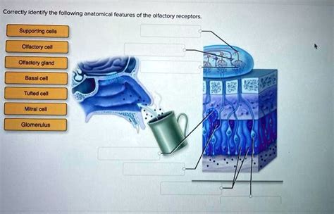
Table of Contents
Correctly Identifying the Anatomical Features of Olfactory Receptors
The sense of smell, or olfaction, is a fascinating and complex process, crucial for survival and deeply intertwined with our memories and emotions. Understanding its mechanics begins with a close examination of the olfactory receptors themselves, the specialized cells responsible for detecting odorants. This article delves into the intricate anatomical features of these receptors, clarifying their structure and function to provide a comprehensive understanding of how we perceive smells.
The Olfactory Epithelium: The Home of Olfactory Receptors
Before we explore the individual receptors, it's crucial to understand their location and environment. Olfactory receptors reside within a specialized region of the nasal cavity called the olfactory epithelium. This patch of tissue, approximately 5cm² in humans, is located high in the nasal cavity, on the superior nasal concha and adjacent regions of the septum. Its unique structure is optimized for efficient odorant detection.
Key Components of the Olfactory Epithelium:
-
Olfactory Receptor Neurons (ORNs): These are the primary sensory neurons responsible for detecting odorants. They are bipolar neurons, meaning they have two processes extending from the cell body: one towards the nasal cavity and the other towards the brain. These are the stars of our show, and we'll explore their detailed anatomy shortly.
-
Supporting Cells: These glial-like cells provide structural and metabolic support to the ORNs, maintaining a healthy environment for their function. They also contribute to the mucus layer.
-
Basal Cells: These are stem cells that continually replace the ORNs, ensuring the olfactory system's ongoing functionality. ORNs have a relatively short lifespan, and basal cells' regenerative capacity is vital for maintaining the sensitivity of smell.
-
Bowman's Glands: Located beneath the olfactory epithelium, these glands secrete mucus that covers the epithelium's surface. This mucus plays a critical role in dissolving odorants, making them accessible to the olfactory receptors. The mucus also contains odorant-binding proteins that help concentrate and transport odorants to the receptor sites.
The Anatomy of Olfactory Receptor Neurons (ORNs): A Detailed Look
Now, let's zoom in on the olfactory receptor neurons themselves, the key players in odor detection. These fascinating cells possess distinct anatomical features that directly contribute to their function.
1. Dendritic Knob and Olfactory Cilia: The Odorant Reception Site
The dendritic knob, a swelling at the end of the ORN's dendrite, projects into the mucus layer of the olfactory epithelium. From this knob, numerous olfactory cilia extend into the mucus. These hair-like structures are the actual sites of odorant reception. Their large surface area significantly increases the chance of odorant molecules interacting with the olfactory receptors.
Importance of Olfactory Cilia:
-
Odorant Receptor Proteins: The cilia's membranes are densely packed with odorant receptor proteins (ORPs). These are G-protein coupled receptors (GPCRs), which means they initiate a signaling cascade upon binding to an odorant molecule. Each ORN expresses only one type of ORP, making them highly specific to certain odorants.
-
High Surface Area: The numerous cilia greatly amplify the surface area available for odorant interaction, increasing the sensitivity of the ORN.
-
Renewal and Regeneration: Olfactory cilia, like the entire ORN, are constantly renewed, ensuring the system’s ongoing sensitivity.
2. Cell Body: The Metabolic Center
The cell body of the ORN, located within the olfactory epithelium, houses the nucleus and other cellular organelles necessary for the cell's metabolic processes. This central region maintains the health and functionality of the entire neuron.
3. Axon: The Signal Transmitter
The axon of the ORN extends from the cell body and passes through the cribriform plate, a bony structure separating the nasal cavity from the cranial cavity. These axons collectively form the olfactory nerve (CN I), which transmits the olfactory signals to the olfactory bulb in the brain. The unique feature of olfactory axons is their ability to regenerate, a testament to the olfactory system's remarkable regenerative capacity.
Odorant Detection and Signal Transduction: A Cellular Perspective
The process of odorant detection involves a precise sequence of events within the olfactory cilia. When an odorant molecule binds to its specific ORP on the cilia, it initiates a cascade of intracellular events leading to the generation of an electrical signal.
The G-Protein Coupled Receptor (GPCR) Pathway:
-
Odorant Binding: An odorant molecule binds to its specific ORP, triggering a conformational change in the receptor.
-
G-protein Activation: This conformational change activates a G-protein, specifically a Golf protein.
-
Adenylate Cyclase Activation: The activated Golf protein stimulates the enzyme adenylate cyclase, increasing the intracellular concentration of cyclic adenosine monophosphate (cAMP).
-
Ion Channel Opening: The increased cAMP binds to and opens cyclic nucleotide-gated (CNG) ion channels in the ciliary membrane, allowing an influx of sodium (Na+) and calcium (Ca2+) ions.
-
Depolarization: The influx of positive ions causes the olfactory cilia to depolarize, generating an electrical signal.
-
Signal Amplification: The process involves multiple amplification steps, ensuring that even a single odorant molecule binding can lead to a significant electrical signal.
The Olfactory Bulb: Relaying the Signal to the Brain
The axons of the olfactory receptor neurons converge to form the olfactory nerve, which projects to the olfactory bulb, a structure in the brain. Here, the signals are processed and relayed to higher brain centers for conscious perception of smell.
Key Features of the Olfactory Bulb:
-
Glomeruli: The olfactory bulb is organized into distinct functional units called glomeruli. Each glomerulus receives input from ORNs expressing the same type of ORP. This organization allows for spatial segregation of olfactory information.
-
Mitral Cells: Within the glomeruli, the olfactory nerve axons synapse with mitral cells, which are the main projection neurons of the olfactory bulb. These cells transmit the processed olfactory information to higher brain regions.
-
Granule Cells: These interneurons in the olfactory bulb play a crucial role in modulating the activity of mitral cells. They contribute to lateral inhibition, sharpening the olfactory signal and improving discrimination between different odors.
Clinical Significance: Olfactory Disorders
Understanding the anatomy of olfactory receptors is crucial for understanding olfactory disorders. Conditions affecting the olfactory epithelium, olfactory nerve, or olfactory bulb can lead to anosmia (loss of smell), hyposmia (reduced smell), or dysosmia (distorted smell). These disorders can be caused by various factors, including:
-
Upper Respiratory Tract Infections: Viral infections can damage the olfactory epithelium, leading to temporary or permanent olfactory loss.
-
Head Trauma: Damage to the cribriform plate can sever olfactory nerve axons, causing anosmia.
-
Neurodegenerative Diseases: Conditions like Parkinson's and Alzheimer's disease are often associated with olfactory dysfunction.
-
Exposure to Toxic Substances: Certain chemicals can damage olfactory receptors, leading to olfactory impairment.
Diagnosis and treatment of olfactory disorders require a detailed understanding of the anatomy and physiology of the olfactory system.
Conclusion: The Intricate World of Olfactory Receptors
The anatomical features of olfactory receptors are intricately designed to facilitate odorant detection and signal transduction. From the specialized cilia to the intricate neural pathways, each component plays a critical role in our ability to perceive smells. This comprehensive understanding not only enhances our appreciation of this vital sensory system but also provides a foundation for further research into olfactory disorders and potential therapeutic interventions. Future research continues to uncover the complexities of olfactory coding and the remarkable plasticity of the olfactory system. This deeper understanding of how we smell will undoubtedly lead to advancements in various fields, from treating olfactory loss to developing novel sensory technologies.
Latest Posts
Latest Posts
Related Post
Thank you for visiting our website which covers about Correctly Identify The Following Anatomical Features Of The Olfactory Receptors. . We hope the information provided has been useful to you. Feel free to contact us if you have any questions or need further assistance. See you next time and don't miss to bookmark.
