Correctly Identify The Following Structures Of The Cochlea
Onlines
Apr 07, 2025 · 6 min read
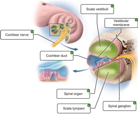
Table of Contents
Correctly Identifying the Structures of the Cochlea: A Comprehensive Guide
The cochlea, a snail-shaped structure residing within the inner ear, plays a pivotal role in our sense of hearing. Its intricate anatomy allows for the transduction of sound vibrations into electrical signals that the brain interprets as sound. Understanding the cochlea's structures is crucial for comprehending the mechanics of hearing and diagnosing auditory disorders. This comprehensive guide delves into the key anatomical components of the cochlea, providing a detailed understanding of their functions and interrelationships.
The Cochlea's Spiral Architecture: An Overview
Before diving into specific structures, it's essential to grasp the overall architecture of the cochlea. Imagine a snail shell uncoiled; this analogy effectively represents the cochlea's spiral shape, comprising approximately 2.5 turns. This spiral structure houses three fluid-filled chambers, crucial for sound transmission and processing. These chambers, separated by membranes, are the scala vestibuli, scala media, and scala tympani.
1. The Scala Vestibuli:
The scala vestibuli is the uppermost chamber of the cochlea. It's connected to the oval window, the entry point for sound vibrations entering the inner ear from the middle ear via the stapes (stirrup bone). The oval window's movement transmits pressure waves into the perilymph, the fluid filling the scala vestibuli and scala tympani. This initiation of pressure waves is the first step in sound transduction.
2. The Scala Media (Cochlear Duct):
Sandwiched between the scala vestibuli and scala tympani, the scala media is a crucial structure, distinct from the other two in its fluid composition and biological importance. It contains endolymph, a fluid with a significantly higher potassium ion concentration than perilymph. This difference in ionic composition is vital for the function of the hair cells, the sensory receptors of hearing. The scala media is also the location of the Organ of Corti, the sensory organ responsible for converting sound vibrations into neural signals.
3. The Scala Tympani:
The scala tympani is the lowermost chamber of the cochlea. It's filled with perilymph, similar to the scala vestibuli. At the apex of the cochlea, the scala vestibuli and scala tympani are connected via the helicotrema, a small opening allowing for the flow of perilymph between the two chambers. This connection is particularly important for low-frequency sound transmission. The scala tympani terminates at the round window, a membrane that releases pressure generated by sound vibrations. The round window's ability to bulge outward is essential for accommodating the pressure changes induced by the sound waves. Without the round window, the pressure wave would be unable to dissipate, significantly impairing hearing.
Key Membranes Within the Cochlea:
Several membranes separate and define the chambers within the cochlea. Understanding their function is crucial for understanding the mechanics of hearing.
1. Reissner's Membrane (Vestibular Membrane):
This thin membrane separates the scala vestibuli from the scala media. While seemingly delicate, Reissner's membrane plays a crucial role in maintaining the ionic difference between the endolymph of the scala media and the perilymph of the scala vestibuli. This ionic difference is vital for the proper functioning of the hair cells. Disruption to Reissner's membrane can compromise this ionic balance, impacting hearing sensitivity.
2. Basilar Membrane:
The basilar membrane forms the floor of the scala media and separates it from the scala tympani. This membrane is not uniform; its width and stiffness vary along its length. This tonotopic organization is fundamental to the cochlea's ability to process sound frequencies. The basilar membrane's base (near the oval window) is narrow and stiff, responding best to high-frequency sounds. Conversely, the basilar membrane's apex (near the helicotrema) is wide and flexible, responding best to low-frequency sounds. This graded response along the basilar membrane allows the cochlea to discern a vast range of sound frequencies. Damage to the basilar membrane, often caused by noise exposure or age-related degeneration, can lead to hearing loss.
The Organ of Corti: The Sensory Epicenter of Hearing
Nestled on the basilar membrane within the scala media lies the Organ of Corti, the sensory organ of hearing. It's a highly structured and complex arrangement of cells responsible for converting mechanical vibrations into electrical signals.
1. Hair Cells: The Transducers of Sound:
The Organ of Corti contains two main types of hair cells: inner hair cells (IHCs) and outer hair cells (OHCs).
-
Inner Hair Cells (IHCs): These are primarily responsible for transmitting auditory information to the brain. They are arranged in a single row and their stereocilia (hair-like structures) are stimulated by the movement of the basilar membrane. This stimulation generates electrical signals that are transmitted to the auditory nerve. The IHCs are the primary sensory receptors for hearing, and their damage results in significant hearing loss.
-
Outer Hair Cells (OHCs): These are arranged in three rows and play a crucial role in amplifying sound vibrations. Their stereocilia are connected to motor proteins that allow them to change length in response to sound. This "cochlear amplifier" mechanism enhances the sensitivity of the basilar membrane, particularly for low-intensity sounds. OHC damage contributes to hearing loss, often resulting in a reduction in sensitivity and clarity of sound.
2. Supporting Cells:
In addition to hair cells, the Organ of Corti contains various supporting cells that provide structural support, nutrition, and ion regulation for the hair cells. These cells play a critical role in maintaining the healthy function of the organ.
3. Tectorial Membrane:
Overlying the hair cells is the tectorial membrane, a gelatinous structure. When the basilar membrane vibrates, the stereocilia of the hair cells are deflected against the tectorial membrane. This deflection opens ion channels in the hair cells, leading to the generation of electrical signals. The interaction between the hair cells and the tectorial membrane is essential for the transduction of sound vibrations into electrical signals.
The Auditory Nerve: Relaying Auditory Information to the Brain
The auditory nerve fibers originate from the spiral ganglion, located beneath the Organ of Corti. These nerve fibers synapse with the hair cells, collecting the electrical signals generated by sound-induced stimulation. These signals are then transmitted to the brainstem, where they undergo further processing before reaching the auditory cortex, the brain region responsible for sound perception. The auditory nerve's integrity is paramount for transmitting accurate auditory information to the brain. Damage to the auditory nerve can result in various types of hearing loss.
Clinical Significance and Diagnostic Imaging:
Understanding the intricate structures of the cochlea is crucial for diagnosing and managing various hearing disorders. Damage to any of these structures can result in different types of hearing loss, ranging from mild to profound. Advanced imaging techniques, such as high-resolution computed tomography (CT) and magnetic resonance imaging (MRI), are utilized to visualize the cochlea and its structures, enabling clinicians to identify abnormalities and guide appropriate interventions.
Conclusion:
The cochlea, with its complex interplay of chambers, membranes, and specialized cells, is a marvel of biological engineering. The precise arrangement of its structures allows for the efficient transduction of sound vibrations into neural signals, enabling our perception of the auditory world. A thorough understanding of the cochlea's anatomy is crucial for appreciating the mechanisms of hearing and for diagnosing and managing hearing disorders. This detailed exploration of the cochlea’s components serves as a foundational resource for anyone seeking to deepen their knowledge of this remarkable sensory organ. Further research and exploration continue to unravel the intricacies of the cochlea and its role in the complex process of hearing. Continued study will further enhance our understanding of this fascinating and vital part of the human auditory system.
Latest Posts
Latest Posts
-
Chapter 19 1 Measuring Recording Height And Weight Answers
Apr 08, 2025
-
A Cuantas Millas De Mexicali Queda Guadalupe Ca
Apr 08, 2025
-
This Textbooks Preferred Model Of Communication Is
Apr 08, 2025
-
Predict The Major Product Of The Reaction Shown
Apr 08, 2025
-
Able Baker And Charles Are Engaged In The Marketing
Apr 08, 2025
Related Post
Thank you for visiting our website which covers about Correctly Identify The Following Structures Of The Cochlea . We hope the information provided has been useful to you. Feel free to contact us if you have any questions or need further assistance. See you next time and don't miss to bookmark.