Correctly Label The Anatomical Features Of Lymphatic Capillaries
Onlines
Mar 16, 2025 · 5 min read
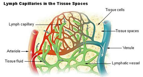
Table of Contents
Correctly Labeling the Anatomical Features of Lymphatic Capillaries
The lymphatic system, often overlooked in discussions of the circulatory system, plays a vital role in maintaining bodily fluid balance, immune function, and overall health. Understanding its intricacies, particularly at the microscopic level of lymphatic capillaries, is crucial for medical professionals and students alike. This article delves deep into the anatomical features of lymphatic capillaries and provides a comprehensive guide for their accurate labeling.
The Importance of Accurate Labeling
Precise labeling of anatomical structures is paramount in medical illustration, education, and research. Inaccurate labeling can lead to misunderstandings, misinterpretations, and even errors in medical practice. For lymphatic capillaries, the correct identification of their unique features is essential for comprehending their function within the broader lymphatic system. This article will equip you with the knowledge to accurately label these delicate vessels and their components.
Identifying Lymphatic Capillaries: A Microscopic Perspective
Lymphatic capillaries, the smallest vessels of the lymphatic system, are microscopic structures that are responsible for the initial uptake of lymph. Unlike blood capillaries, which form a continuous network, lymphatic capillaries are characterized by their unique structure that facilitates fluid absorption. Their features are distinctly different and necessitate careful observation under a microscope.
Key Anatomical Features and Their Labeling
-
Blind-Ended Nature: Unlike blood capillaries, which form continuous loops, lymphatic capillaries are blind-ended. This means they have one open end, allowing interstitial fluid to enter but not exit directly back into the same capillary. This is crucial for the unidirectional flow of lymph. When labeling, clearly indicate the closed end versus the open end of the capillary.
-
Overlapping Endothelial Cells: The walls of lymphatic capillaries are composed of a single layer of endothelial cells. These cells are uniquely arranged, overlapping in a manner that creates one-way valves. These valves prevent backflow of lymph and ensure its movement towards larger lymphatic vessels. Labeling should clearly depict this overlap, highlighting how this unique arrangement contributes to the valve-like function.
-
Anchoring Filaments: Extending from the endothelial cells are thin, filamentous structures called anchoring filaments. These filaments attach the lymphatic capillary wall to the surrounding connective tissue. When interstitial fluid pressure increases, these filaments pull on the endothelial cells, widening the gaps between them. This facilitates the entry of fluid, proteins, and even larger particles, such as cellular debris and bacteria, into the lymphatic capillary. Label these anchoring filaments and describe their role in facilitating lymph uptake.
-
Intercellular Junctions: The endothelial cells are connected by intercellular junctions. These junctions are not as tight as those in blood capillaries, creating relatively large gaps that allow for the passage of larger molecules and even cells. These junctions are crucial for the lymphatic system's role in immune surveillance and the transport of immune cells. Clearly label these loose junctions to emphasize their permeability.
-
Lymph: The fluid within the lymphatic capillary is called lymph. It’s a clear to pale yellow fluid containing water, proteins, fats, and various immune cells. While not a structural feature of the capillary itself, it’s vital to show lymph within the lumen of the lymphatic capillary during labeling. Highlighting its composition is beneficial for understanding lymphatic function.
-
Basement Membrane (Often Incomplete): Unlike blood capillaries, lymphatic capillaries may possess an incomplete or discontinuous basement membrane. This contributes to their increased permeability and ability to absorb larger molecules and cells. Labeling this feature should reflect its incompleteness compared to the continuous basement membrane found in blood capillaries.
-
Surrounding Connective Tissue: Lymphatic capillaries are embedded within the surrounding connective tissue, which provides structural support and facilitates interaction with other tissues and cells. Include the connective tissue in your labeling, emphasizing its role in anchoring the capillaries and contributing to the overall microenvironment.
Illustrating Lymphatic Capillaries: Tips for Accurate Representation
Creating clear and accurate illustrations of lymphatic capillaries requires attention to detail and adherence to the known anatomy. Consider the following tips:
-
Use appropriate magnification: Microscopic features like overlapping endothelial cells and anchoring filaments are best depicted using a high magnification.
-
Employ clear labeling: Use clear, concise labels with consistent font and size. Avoid cluttered diagrams; less is often more effective.
-
Illustrate the 3D structure: Lymphatic capillaries are not flat structures. Attempt to show their three-dimensional arrangement, including how they weave through the tissues.
-
Use color-coding: Consider using different colors to differentiate between different structural components (e.g., endothelial cells, anchoring filaments, lymph).
-
Include a scale bar: Provide a scale bar to indicate the size of the structures shown in your illustration.
Clinical Significance of Lymphatic Capillaries
Understanding the anatomy of lymphatic capillaries has significant clinical implications. Their dysfunction can lead to various conditions, including:
-
Lymphedema: A condition characterized by swelling due to impaired lymphatic drainage.
-
Lipedema: A disorder that primarily affects the legs and involves abnormal fat accumulation.
-
Cancer metastasis: Cancer cells can utilize lymphatic capillaries to spread to other parts of the body.
-
Infectious diseases: Lymphatic capillaries play a critical role in immune response to infection.
Accurate labeling and understanding of lymphatic capillary anatomy are vital in diagnosing and treating these conditions.
Conclusion: Mastery Through Accurate Labeling
Mastering the accurate labeling of lymphatic capillary anatomy requires careful study and attention to detail. By understanding the unique features of these vessels, including their blind-ended nature, overlapping endothelial cells, anchoring filaments, and incomplete basement membrane, you can create clear, accurate illustrations and contribute to a deeper understanding of this crucial component of the lymphatic system. This detailed knowledge empowers medical professionals to better understand the implications of lymphatic dysfunction and develop more effective diagnostic and therapeutic strategies. Consistent and correct labeling is the cornerstone of effective communication and a fundamental skill for anyone working in the field of anatomy, physiology, or medicine. The accuracy of your labeling directly impacts the understanding and interpretation of the complex biological processes taking place at this microscopic level.
Latest Posts
Latest Posts
-
Add The Text Slow Start To The Shape
Mar 16, 2025
-
No Portion Of This Ad Shows What This Thing Does
Mar 16, 2025
-
How Much Does A Sandwich Bag Weigh
Mar 16, 2025
-
Which Of The Following Statements Does Not Describe Brucellosis
Mar 16, 2025
-
A Factor That Causes Overhead Costs Is Called A Blank
Mar 16, 2025
Related Post
Thank you for visiting our website which covers about Correctly Label The Anatomical Features Of Lymphatic Capillaries . We hope the information provided has been useful to you. Feel free to contact us if you have any questions or need further assistance. See you next time and don't miss to bookmark.
