Correctly Label The Following Anatomical Features Of The Cerebellum.
Onlines
Mar 13, 2025 · 7 min read
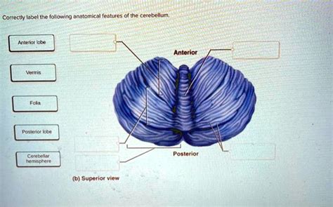
Table of Contents
Correctly Label the Anatomical Features of the Cerebellum: A Comprehensive Guide
The cerebellum, often called the "little brain," is a fascinating and vital structure nestled at the base of the brain. While smaller than the cerebrum, its role in coordinating movement, balance, and posture is absolutely critical. Understanding its intricate anatomy is crucial for anyone studying neuroscience, neurology, or related fields. This comprehensive guide will delve into the key anatomical features of the cerebellum, providing detailed descriptions and assisting you in correctly labeling them.
I. Gross Anatomy: External Features of the Cerebellum
Before diving into the microscopic details, let's establish a firm understanding of the cerebellum's overall structure. Its external appearance is distinctive, characterized by several key features:
1. Cerebellar Hemispheres:
These two large, lateral lobes dominate the cerebellum's appearance. They are separated by a deep fissure, the vermis, and each hemisphere is further subdivided into lobes. The surface of the hemispheres, like the cerebrum, is highly convoluted, increasing surface area for neuronal processing. This convoluted surface is characterized by numerous folia, which are thin, parallel folds. These are analogous to the gyri found in the cerebrum.
2. Vermis:
The vermis is the central, unpaired portion of the cerebellum. It's a narrow band of tissue that runs along the midline, connecting the two cerebellar hemispheres. The vermis plays a crucial role in mediating axial motor control, which governs movements of the trunk and proximal limbs. It integrates sensory information from various parts of the body to coordinate these movements. Its location and function make it a central hub for cerebellar processing.
3. Lobes of the Cerebellum:
While the boundaries are not always sharply defined, the cerebellum is often broadly divided into lobes:
- Anterior Lobe: Situated anteriorly, this lobe is primarily involved in the regulation of muscle tone and the coordination of posture and locomotion.
- Posterior Lobe: Occupying the majority of the cerebellar surface, this lobe is concerned with the fine-tuning and coordination of skilled voluntary movements. Damage to this lobe often results in ataxia (lack of coordination).
- Flocculonodular Lobe: This lobe, situated inferiorly, plays a key role in maintaining equilibrium and balance. It receives input from the vestibular system, which is responsible for sensing head position and movement.
4. Cerebellar Peduncles:
These are three pairs of thick fiber bundles that connect the cerebellum to the brainstem. They are essential for communication between the cerebellum and other parts of the nervous system:
- Superior Cerebellar Peduncles: Primarily efferent (carrying signals away from the cerebellum), they carry signals to the midbrain and thalamus.
- Middle Cerebellar Peduncles: Primarily afferent (carrying signals towards the cerebellum), they receive input from the pontine nuclei.
- Inferior Cerebellar Peduncles: Carry both afferent and efferent fibers. Afferent fibers bring sensory information from the spinal cord and vestibular nuclei, while efferent fibers project to the vestibular nuclei and reticular formation.
II. Internal Anatomy: Deeper Structures of the Cerebellum
Looking beyond the surface, the internal organization of the cerebellum is equally complex and crucial to its function.
1. White Matter:
The internal core of the cerebellum consists of white matter, composed primarily of myelinated axons. This white matter forms a branched, tree-like structure known as the arbor vitae, meaning "tree of life," due to its distinctive appearance in cross-section. This intricate network of axons facilitates the rapid transmission of information throughout the cerebellum.
2. Deep Cerebellar Nuclei:
Embedded within the white matter are four pairs of deep cerebellar nuclei, which play a vital role in relaying cerebellar output:
- Dentate Nucleus: The largest of the deep cerebellar nuclei, the dentate nucleus is involved in the planning and execution of complex voluntary movements. It projects primarily to the ventrolateral thalamus.
- Emboliform Nucleus: Situated medial to the dentate nucleus, this nucleus is implicated in the coordination of limb movements. It also projects to the thalamus.
- Globose Nucleus: Located medial to the emboliform nucleus, this nucleus is involved in similar functions to the emboliform nucleus, contributing to the coordination of movement. It also projects to the thalamus.
- Fastigial Nucleus: The most medially located nucleus, the fastigial nucleus is predominantly involved in the regulation of posture, balance, and eye movements. It projects to the vestibular nuclei and reticular formation.
3. Cerebellar Cortex:
The outer layer of the cerebellum is the cerebellar cortex, a highly organized structure responsible for the complex processing of sensory and motor information. It is composed of three layers:
- Molecular Layer: The outermost layer, it contains relatively few neurons but a dense network of axons and dendrites, along with interneurons like stellate and basket cells.
- Purkinje Cell Layer: This single layer contains the large, distinctive Purkinje cells, which are the only output neurons of the cerebellar cortex. Their extensive dendritic arborizations receive input from granule cells and climbing fibers.
- Granular Layer: The innermost layer, it is packed with densely arranged granule cells, the most numerous neurons in the brain. These granule cells receive input from mossy fibers and their axons ascend to the molecular layer, where they bifurcate to form parallel fibers.
III. Functional Anatomy: Cerebellar Circuits and Pathways
The cerebellum's sophisticated structure underpins its vital functions. Understanding the flow of information through cerebellar circuits is key:
1. Mossy Fiber Input:
Mossy fibers originate from various sources, including the spinal cord, brainstem, and pontine nuclei. They carry sensory and motor information to the cerebellar cortex, synapsing on granule cells in the granular layer.
2. Climbing Fiber Input:
Climbing fibers originate from the inferior olivary nucleus in the medulla. They are characterized by their strong excitatory effect on Purkinje cells, which is essential for motor learning and adaptation. Each Purkinje cell receives input from only one climbing fiber.
3. Purkinje Cell Output:
Purkinje cells are the sole output neurons of the cerebellar cortex. They inhibit the deep cerebellar nuclei via GABAergic synapses. This inhibitory effect plays a crucial role in shaping the motor commands that are relayed to other brain areas.
4. Deep Nuclei Output:
The deep cerebellar nuclei receive inhibitory input from Purkinje cells and excitatory input from mossy and climbing fibers. They then project to various areas of the brain, including the thalamus, brainstem, and vestibular nuclei, influencing motor control, balance, and coordination.
IV. Clinical Significance: Cerebellar Dysfunction
Damage or dysfunction of the cerebellum can have significant consequences, resulting in a range of neurological disorders:
1. Ataxia:
This is the most prominent symptom of cerebellar damage. Ataxia manifests as a lack of coordination of voluntary movements, often resulting in jerky, inaccurate movements and difficulty with balance.
2. Dysmetria:
This refers to an inability to accurately judge distances, leading to overshooting or undershooting movements. It is commonly observed in cerebellar lesions.
3. Tremor:
Intentional tremors, which worsen during voluntary movements, are frequently associated with cerebellar damage. These tremors are distinct from resting tremors seen in Parkinson's disease.
4. Nystagmus:
This refers to involuntary rhythmic oscillations of the eyes, often indicative of cerebellar dysfunction, especially lesions in the flocculonodular lobe.
5. Hypotonia:
Reduced muscle tone is another potential consequence of cerebellar damage, leading to weakness and diminished resistance to passive movement.
Understanding the cerebellum's intricate anatomy and functional pathways is crucial for clinicians to diagnose and manage cerebellar disorders effectively. Accurate identification of specific regions of the cerebellum affected by pathology can aid in prognosis and guide therapeutic interventions.
V. Labeling Practice: A Step-by-Step Approach
To reinforce your understanding, let's practice labeling the key anatomical features. Imagine a diagram of the cerebellum:
-
Identify the Cerebellar Hemispheres: Locate the two large lateral lobes.
-
Locate the Vermis: Identify the central, unpaired structure separating the hemispheres.
-
Distinguish the Lobes: Try to differentiate the anterior, posterior, and flocculonodular lobes. Remember these boundaries are not always sharply defined.
-
Pinpoint the Cerebellar Peduncles: Locate the three pairs of fiber bundles connecting the cerebellum to the brainstem.
-
Visualize the Arbor Vitae: Imagine the branching pattern of the white matter within the cerebellum.
-
Identify the Deep Cerebellar Nuclei: Locate the four pairs of nuclei embedded within the white matter: dentate, emboliform, globose, and fastigial.
-
Recognize the Cerebellar Cortex Layers: Mentally separate the molecular, Purkinje cell, and granular layers.
By systematically reviewing these structures and their relationships, you'll build a strong foundation for understanding the cerebellum's complex anatomy. Regular practice with anatomical diagrams and models will further enhance your ability to accurately label and comprehend this critical brain structure. The more you engage with this material, the more proficient you will become in recognizing and correctly identifying each component. Remember, mastering the cerebellum's anatomy is a journey, not a race, and consistent effort will lead to success.
Latest Posts
Latest Posts
-
Ap Lit Unit 2 Progress Check Mcq
Mar 13, 2025
-
Simulation Lab 15 1 Module 15 Using A Nonpersistent Web Browser
Mar 13, 2025
-
Geometry Basics Unit 1 Homework 1
Mar 13, 2025
-
The Spirit Catches Me And I Fall Down Sparknotes
Mar 13, 2025
-
Table 19 1 Summary Table Of Animal Characteristics
Mar 13, 2025
Related Post
Thank you for visiting our website which covers about Correctly Label The Following Anatomical Features Of The Cerebellum. . We hope the information provided has been useful to you. Feel free to contact us if you have any questions or need further assistance. See you next time and don't miss to bookmark.
