Correctly Label The Following Anatomical Features Of The Spinal Cord
Onlines
Mar 10, 2025 · 7 min read
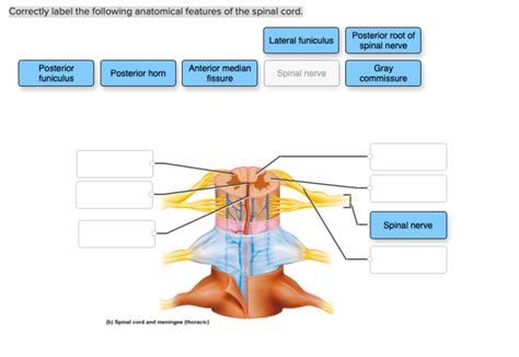
Table of Contents
Correctly Labeling the Anatomical Features of the Spinal Cord: A Comprehensive Guide
The spinal cord, a crucial component of the central nervous system, is a fascinating and complex structure. Understanding its intricate anatomy is essential for anyone studying biology, medicine, or related fields. This comprehensive guide will delve into the key anatomical features of the spinal cord, providing detailed descriptions and visual aids to help you correctly label them. We'll cover everything from external features to internal structures, ensuring you gain a thorough understanding of this vital organ.
External Anatomy of the Spinal Cord: A Visual Overview
Before delving into the microscopic details, let's familiarize ourselves with the spinal cord's external features. Imagine a long, cylindrical structure extending from the medulla oblongata (the lower part of the brainstem) to approximately the level of the first lumbar vertebra (L1). This is the spinal cord, protected within the vertebral canal of the spine.
Key External Features to Identify:
-
Cervical Enlargement: Notice the slightly thicker region in the cervical (neck) area of the spinal cord. This enlargement, superior to the thoracic region, is crucial because it houses the nerve roots that innervate the upper limbs. The increased size reflects the greater number of neurons needed to control these limbs' complex movements.
-
Lumbosacral Enlargement: Similarly, observe the other prominent enlargement located in the lumbar region (lower back). This area is responsible for innervating the lower limbs, explaining its increased size compared to the thoracic region.
-
Conus Medullaris: The spinal cord doesn't extend the entire length of the vertebral column. Instead, it tapers to a cone-shaped end called the conus medullaris, usually ending around the L1 vertebra.
-
Filum Terminale: This is a delicate, fibrous extension that continues from the conus medullaris. It anchors the spinal cord to the coccyx, providing crucial structural support and stability.
-
Cauda Equina: Below the conus medullaris, the nerve roots of the lumbar, sacral, and coccygeal segments extend inferiorly within the vertebral canal, resembling a horse's tail. This is the cauda equina, an important structure because its nerves supply the lower limbs and pelvic organs. The individual nerves within the cauda equina are significantly longer than those in the upper spinal cord segments due to the difference in vertebral length and spinal cord termination point.
-
Anterior Median Fissure: This is a deep, longitudinal groove on the anterior (front) surface of the spinal cord. It is a significant landmark for identifying the anterior aspect.
-
Posterior Median Sulcus: On the posterior (back) surface, find a shallower, less prominent groove called the posterior median sulcus. It marks the midline of the posterior aspect of the spinal cord.
-
Dorsal Rootlets and Dorsal Roots: Emerging from the posterior aspect of the spinal cord are several small rootlets that merge to form the dorsal (posterior) root of each spinal nerve. These roots carry sensory information from the periphery to the spinal cord.
-
Ventral Rootlets and Ventral Roots: From the anterior aspect of the spinal cord, ventral (anterior) rootlets emerge. These fuse to form the ventral root, containing motor axons that carry signals from the spinal cord to muscles and glands.
-
Spinal Nerve: Finally, the dorsal and ventral roots of each segment unite to form the spinal nerve, which carries both sensory and motor fibers. This marks the crucial point where sensory and motor information converge and diverge.
Internal Anatomy: Unveiling the Spinal Cord's Microscopic World
The external anatomy provides a roadmap, but the real magic lies within. Let's explore the internal structures, which are critical to understanding the spinal cord's function.
Cross-Sectional View: A Detailed Look Inside
A cross-section of the spinal cord reveals two main regions:
-
Gray Matter: The butterfly-shaped area in the center is the gray matter. It is predominantly composed of neuronal cell bodies, dendrites, and unmyelinated axons, giving it a darker appearance.
-
White Matter: Surrounding the gray matter is the white matter, appearing lighter due to the presence of myelinated axons. These axons transmit signals up and down the spinal cord, facilitating communication between different parts of the nervous system.
Within the Gray Matter: Key Structures
The gray matter's "wings" are known as horns, and each has specific functions:
-
Posterior Horns (Dorsal Horns): These horns receive sensory information carried by the dorsal roots. The cells within the posterior horns process sensory input before relaying it to the brain or other parts of the spinal cord. Sensory neurons primarily reside within the dorsal root ganglia (which are outside the spinal cord itself, but closely associated).
-
Anterior Horns (Ventral Horns): These horns contain the cell bodies of motor neurons, whose axons extend out through the ventral roots to innervate skeletal muscles. The anterior horns are responsible for the motor commands responsible for voluntary muscle movements.
-
Lateral Horns: Found only in the thoracic and upper lumbar regions of the spinal cord, the lateral horns contain the cell bodies of autonomic neurons that regulate involuntary functions like heartbeat and digestion. Their functions differ from the anterior and posterior horns, highlighting the specialized organization of the spinal cord.
Within the White Matter: Tracts and Pathways
The white matter consists of several tracts, bundles of myelinated axons that run in specific directions:
-
Ascending Tracts: These carry sensory information from the body to the brain. Examples include the spinothalamic tract (transmitting pain and temperature), dorsal column-medial lemniscus pathway (carrying touch and proprioception information), and spinocerebellar tracts (conveying proprioceptive information to the cerebellum).
-
Descending Tracts: These tracts carry motor commands from the brain to the body. The corticospinal tract, crucial for voluntary motor control, is a prominent example. Other descending tracts control posture, balance, and autonomic functions.
Clinical Significance: Understanding Spinal Cord Injuries and Diseases
Understanding the spinal cord's anatomy is critical for diagnosing and treating various neurological conditions. Damage to specific areas can result in diverse symptoms, highlighting the importance of anatomical precision.
Examples of Clinical Relevance:
-
Spinal Cord Injury: Trauma to the spinal cord can lead to paralysis, loss of sensation, and other debilitating effects. The severity and location of the injury determine the specific symptoms. For example, damage to the cervical spinal cord might lead to quadriplegia (paralysis of all four limbs), whereas damage to the thoracic region could cause paraplegia (paralysis of the lower body).
-
Multiple Sclerosis (MS): This autoimmune disease attacks the myelin sheath surrounding axons in the central nervous system, including the spinal cord. This demyelination disrupts the transmission of nerve impulses, leading to various neurological symptoms.
-
Amyotrophic Lateral Sclerosis (ALS): Also known as Lou Gehrig's disease, ALS is a progressive neurodegenerative disease that affects motor neurons in the brain and spinal cord. It leads to muscle weakness, atrophy, and ultimately paralysis.
-
Spinal Muscular Atrophy (SMA): This group of genetic disorders affects motor neurons, resulting in muscle weakness and atrophy. The severity varies depending on the specific type of SMA.
Accurate labeling of the spinal cord's anatomical features is essential for understanding its function in health and disease. Clinicians rely on this knowledge to diagnose and manage a wide range of neurological disorders. Detailed anatomical understanding is crucial for proper assessment and treatment of any spinal cord pathology.
Practical Application: Labeling Exercises and Resources
To solidify your understanding, practice labeling diagrams of the spinal cord. You can find numerous resources online, in textbooks, and anatomy atlases. Focus on identifying the structures mentioned above, ensuring you can distinguish between the anterior and posterior aspects, gray and white matter, and the various tracts and horns.
Regular practice and reinforcement will improve your understanding of the complex anatomy of the spinal cord. Remember, mastery of this subject requires consistent effort and engagement with various learning tools and methodologies.
Conclusion: Mastering the Anatomy of the Spinal Cord
Correctly labeling the anatomical features of the spinal cord requires a thorough understanding of its external and internal structures. This guide has provided a detailed overview, equipping you with the knowledge to accurately identify and label key anatomical features. By understanding the function of each component and its clinical significance, you'll develop a strong foundation in neuroanatomy. Remember that continued practice and engagement with visual aids will be invaluable in mastering this complex yet fascinating subject. This comprehensive knowledge will be instrumental for further studies in biology, medicine, and related fields.
Latest Posts
Latest Posts
-
The Tragedy Of Romeo And Juliet Act 2 Cloze Activity
Mar 11, 2025
-
A Feast For Crows Plot Summary
Mar 11, 2025
-
Jasmin Belongs To The Chess Club On Her Campus
Mar 11, 2025
Related Post
Thank you for visiting our website which covers about Correctly Label The Following Anatomical Features Of The Spinal Cord . We hope the information provided has been useful to you. Feel free to contact us if you have any questions or need further assistance. See you next time and don't miss to bookmark.
