Correctly Label The Following Anatomical Features Of The Talocrural Joint
Onlines
Mar 23, 2025 · 6 min read
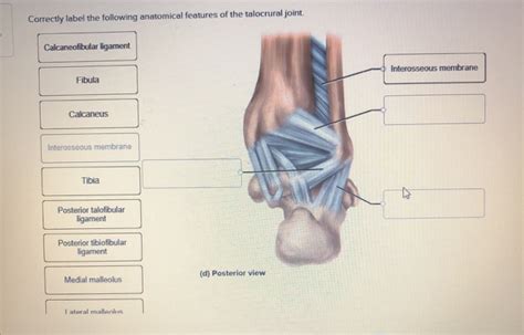
Table of Contents
Correctly Labeling the Anatomical Features of the Talocrural Joint
The talocrural joint, also known as the ankle joint, is a crucial articulation responsible for the complex movements of the foot and ankle. Understanding its intricate anatomy is paramount for clinicians, students, and anyone interested in the biomechanics of lower limb locomotion. This comprehensive guide will delve into the detailed anatomy of the talocrural joint, focusing on the correct labeling of its key features. We will explore the bones involved, the ligaments that provide stability, and the surrounding musculature that contributes to its dynamic function.
The Bones of the Talocrural Joint
The talocrural joint is a modified hinge joint, formed by the articulation of three bones:
1. Tibia: The Major Weight-Bearing Bone
The tibia, or shin bone, is the larger and medially located of the two lower leg bones. Its distal end, specifically the tibial plafond, plays a crucial role in forming the superior aspect of the talocrural joint. The medial malleolus, a prominent bony projection on the medial side of the distal tibia, contributes significantly to the joint stability by articulating with the talus. The articular surface of the tibia is relatively flat, providing a stable base for the talus. Understanding the tibial plafond's shape and congruity with the talus is crucial for comprehending normal ankle mechanics and diagnosing various ankle pathologies.
2. Fibula: Lateral Stability
The fibula, the thinner lateral bone of the lower leg, also contributes to the talocrural joint, albeit indirectly. Its distal end, the lateral malleolus, forms the lateral wall of the ankle mortise. While the fibula doesn't directly articulate with the talus to the same extent as the tibia, its lateral malleolus is essential for maintaining the joint's structural integrity and lateral stability. The strong ligamentous connections between the fibula and the talus and calcaneus further highlight the fibula's critical role in ankle stability. Fractures of the lateral malleolus are common ankle injuries, often resulting in instability and subsequent complications.
3. Talus: The Keystone of the Ankle
The talus, a unique bone located in the midfoot, is the keystone of the ankle joint. Its superior surface, the trochlear surface, articulates with the tibial plafond, forming the main weight-bearing surface of the talocrural joint. The trochlear surface is shaped like a spool, allowing for dorsiflexion and plantarflexion. The talus also articulates with the calcaneus (heel bone) inferiorly, and the navicular bone anteriorly, forming the subtalar and talonavicular joints, respectively. The talus's unique shape and articulation with multiple bones contribute to the complex movement patterns of the ankle and foot. The medial and lateral articular facets of the talus are crucial for proper alignment and movement within the ankle mortise. Damage or incongruity in these facets can lead to significant functional limitations.
The Ligaments of the Talocrural Joint: Guardians of Stability
Several strong ligaments reinforce the talocrural joint, providing crucial stability against various stresses. Understanding their precise location and function is crucial for diagnosing and treating ankle injuries:
1. Deltoid Ligament: Medial Stability
The deltoid ligament is a strong, triangular ligament located on the medial aspect of the ankle. It comprises four distinct components:
- Tibionavicular part: Connects the medial malleolus of the tibia to the navicular bone.
- Tibiocalcaneal part: Connects the medial malleolus to the sustentaculum tali of the calcaneus.
- Tibiotalar anterior part: Connects the medial malleolus to the talus.
- Tibiotalar posterior part: Connects the medial malleolus to the talus.
Collectively, these components provide significant medial stability, resisting eversion forces that could lead to ankle sprains or fractures. Injuries to the deltoid ligament are less common than lateral ankle sprains, but they can result in significant instability.
2. Lateral Collateral Ligaments: Lateral Support
The lateral aspect of the ankle is stabilized by three distinct ligaments:
- Anterior talofibular ligament (ATFL): The most commonly injured ligament in lateral ankle sprains. It connects the anterior aspect of the lateral malleolus to the anterior talus.
- Calcaneofibular ligament (CFL): Connects the lateral malleolus to the calcaneus.
- Posterior talofibular ligament (PTFL): The strongest of the three, connecting the posterior aspect of the lateral malleolus to the posterior talus. It is less frequently injured than the ATFL and CFL.
Understanding the relative roles of these ligaments is crucial for diagnosing the grade of a lateral ankle sprain, which is classified based on the number and severity of ligamentous injuries.
The Ankle Mortise: The Functional Joint Space
The ankle mortise is the crucial bony structure formed by the articulation of the distal tibia and fibula with the talus. It's a vital component of the talocrural joint, creating a stable and congruent space for the talus to move within. The precise alignment and congruity of the mortise are critical for maintaining normal ankle mechanics. Any disruption, such as a fracture of the malleoli or ligamentous injury, can lead to malalignment and instability. The integrity of the ankle mortise is essential for weight-bearing and efficient locomotion.
Musculature Contributing to Talocrural Joint Movement
While not directly part of the joint structure, surrounding muscles play a vital role in the movement and stability of the talocrural joint:
- Dorsiflexion: This movement, bringing the toes towards the shin, is primarily facilitated by the tibialis anterior, extensor hallucis longus, extensor digitorum longus, and peroneus tertius muscles.
- Plantarflexion: This movement, pointing the toes downwards, is primarily achieved by the gastrocnemius, soleus, tibialis posterior, peroneus longus, peroneus brevis, and flexor hallucis longus and flexor digitorum longus muscles. These muscles work synergistically to control plantar flexion and provide stability during weight-bearing activities.
Clinical Significance: Understanding Common Injuries
Correctly labeling the anatomical features of the talocrural joint is essential for accurate diagnosis and management of ankle injuries. Common injuries include:
- Lateral Ankle Sprains: These are the most common ankle injuries, often involving injury to the ATFL, CFL, and sometimes the PTFL. The severity of the sprain is classified based on the extent of ligamentous damage.
- Medial Ankle Sprains: Less common than lateral sprains, these injuries involve the deltoid ligament and are often associated with other fractures.
- Ankle Fractures: These can involve the distal tibia, fibula, or talus and often require surgical intervention. Understanding the precise location and type of fracture is crucial for appropriate treatment.
- Osteoarthritis: Degenerative changes in the articular cartilage of the talocrural joint can lead to pain, stiffness, and reduced range of motion.
Conclusion: The Importance of Precise Labeling
Precise labeling of the anatomical features of the talocrural joint is fundamental for accurate communication among healthcare professionals, for effective teaching and learning, and for a thorough understanding of ankle biomechanics. This knowledge is crucial for diagnosing and managing a wide range of ankle injuries and conditions. A comprehensive understanding of the bones, ligaments, and muscles involved is essential for clinicians, researchers, and students alike. By mastering the precise labeling of these structures, we can better understand the complexity and importance of this vital joint in the human body. Continued study and refinement of our knowledge base are crucial for improving the diagnosis, treatment, and prevention of ankle injuries. This will lead to better patient outcomes and a greater understanding of the intricate mechanics of human movement.
Latest Posts
Latest Posts
-
Before Creating A Product It Is Wise To
Mar 23, 2025
-
Chapter 5 Supply Practice Worksheet Answers
Mar 23, 2025
-
Busi 3200 Professional Development Iii Unt
Mar 23, 2025
-
Summary Of The Lesson Toni Cade Bambara
Mar 23, 2025
-
Chapter 23 To Kill A Mockingbird Summary
Mar 23, 2025
Related Post
Thank you for visiting our website which covers about Correctly Label The Following Anatomical Features Of The Talocrural Joint . We hope the information provided has been useful to you. Feel free to contact us if you have any questions or need further assistance. See you next time and don't miss to bookmark.
