Correctly Label The Following Muscles Of The Posterior View
Onlines
Mar 22, 2025 · 6 min read
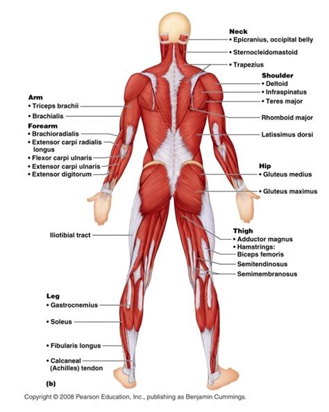
Table of Contents
Correctly Labeling the Muscles of the Posterior View: A Comprehensive Guide
The posterior view of the human body reveals a complex network of muscles responsible for movement, posture, and stability. Correctly identifying these muscles is crucial for anyone studying anatomy, kinesiology, or pursuing a career in healthcare. This comprehensive guide will delve into the detailed labeling of the posterior muscles, providing a thorough understanding of their location, function, and clinical significance. We’ll cover everything from superficial to deep muscles, ensuring a complete understanding of this intricate anatomical region.
Superficial Muscles of the Posterior View
The superficial muscles are the most readily visible and easily palpated. Understanding these forms a strong foundation for identifying the deeper muscle layers.
1. Trapezius:
The trapezius is a large, flat, triangular muscle covering the upper back and neck. It’s easily recognizable by its broad shape and extensive attachments.
- Origin: Occipital bone, ligamentum nuchae, and spinous processes of C7-T12 vertebrae.
- Insertion: Lateral third of the clavicle, acromion process, and spine of the scapula.
- Function: Elevation, retraction, depression, and upward rotation of the scapula. It also extends the head and neck.
- Clinical Significance: Trapezius strain is common, often caused by repetitive movements or poor posture. Weakness can lead to shoulder instability and impaired movement.
2. Latissimus Dorsi:
The latissimus dorsi ("lats") is a large, broad muscle covering the lower back. Its shape resembles a V, with its apex pointing towards the axilla.
- Origin: Spinous processes of T7-L5 vertebrae, thoracolumbar fascia, iliac crest, and inferior three or four ribs.
- Insertion: Intertubercular groove of the humerus.
- Function: Extension, adduction, and medial rotation of the humerus. It also contributes to respiration.
- Clinical Significance: Latissimus dorsi injuries are possible from forceful movements or overstretching. Weakness can impact arm strength and stability.
3. Deltoid:
Although partially visible in the posterior view, the posterior fibers of the deltoid muscle are significant.
- Origin: Lateral third of the clavicle, acromion process, and spine of the scapula.
- Insertion: Deltoid tuberosity of the humerus.
- Function: Posterior fibers extend, laterally rotate, and horizontally abduct the humerus.
- Clinical Significance: Deltoid injuries, such as tears or strains, are common in athletes involved in overhead activities.
4. Levator Scapulae:
A smaller muscle located deep to the trapezius, the levator scapulae is involved in scapular movement.
- Origin: Transverse processes of C1-C4 vertebrae.
- Insertion: Medial border of the scapula, superior to the spine.
- Function: Elevation and downward rotation of the scapula. It also contributes to neck flexion.
- Clinical Significance: Levator scapulae tightness can contribute to neck pain and headaches.
5. Rhomboid Major and Minor:
These two muscles lie deep to the trapezius and work together to stabilize the scapula.
- Origin: Rhomboid major: spinous processes of T2-T5 vertebrae. Rhomboid minor: spinous processes of C7-T1 vertebrae.
- Insertion: Medial border of the scapula.
- Function: Retraction and downward rotation of the scapula.
- Clinical Significance: Weakness or tightness can lead to scapular winging and poor posture.
Deep Muscles of the Posterior View
The deeper muscles are less visible but play crucial roles in posture, stability, and fine motor control. Accessing and understanding these muscles requires a more detailed anatomical knowledge.
6. Erector Spinae Muscle Group:
This group comprises three columns of muscles running vertically along the spine: iliocostalis, longissimus, and spinalis.
- Origin: Iliac crest, sacrum, and spinous processes of lumbar vertebrae.
- Insertion: Ribs, transverse processes of vertebrae, and occipital bone.
- Function: Extension of the vertebral column, lateral flexion, and rotation. These muscles are critical for maintaining posture and stability.
- Clinical Significance: Sprains and strains of the erector spinae muscles are common causes of lower back pain.
7. Quadratus Lumborum:
Located on each side of the lumbar spine, the quadratus lumborum is a deep muscle involved in lateral flexion and stabilization.
- Origin: Iliac crest and iliolumbar ligament.
- Insertion: Transverse processes of L1-L4 vertebrae and 12th rib.
- Function: Lateral flexion of the trunk, extension of the lumbar spine, and stabilization of the pelvis.
- Clinical Significance: Strains or spasms of the quadratus lumborum can cause lower back pain and restricted movement.
8. Multifidus:
This group of small muscles lies deep to the erector spinae and is crucial for spinal stability.
- Origin: Sacrum, posterior iliac crest, and transverse processes of lumbar, thoracic, and cervical vertebrae.
- Insertion: Spinous processes of vertebrae superior to their origin.
- Function: Extension, rotation, and stabilization of the vertebral column.
- Clinical Significance: Weakness in the multifidus muscles is often associated with lower back pain and instability.
9. Deep Intrinsic Back Muscles:
Several smaller, deep muscles, such as the semispinalis, rotatores, and interspinales, contribute to fine motor control of the vertebral column. They are difficult to isolate and palpate individually.
- Origin and Insertion: Vary depending on the specific muscle, generally involving the transverse and spinous processes of the vertebrae.
- Function: Extension, rotation, and stabilization of the vertebral column. Their coordinated actions contribute to precise movements and postural adjustments.
- Clinical Significance: These muscles are often implicated in chronic back pain and may be affected by injury or postural imbalances.
Clinical Correlations and Importance of Correct Labeling
Accurate labeling of the posterior muscles is vital for several reasons:
-
Diagnosis and Treatment: Healthcare professionals rely on precise anatomical knowledge to diagnose injuries, assess dysfunction, and develop effective treatment plans. Mislabeling muscles can lead to misdiagnosis and inappropriate therapies.
-
Physical Therapy: Physical therapists utilize their understanding of muscle anatomy to design targeted exercises for rehabilitation and injury prevention. Precise labeling allows them to create effective treatment programs.
-
Sports Medicine: Athletes often experience injuries to posterior muscles. Correct identification is essential for accurate diagnosis, management, and return-to-play decisions.
-
Surgical Procedures: Surgeons require a detailed understanding of the anatomy of the back to avoid damaging structures during surgical interventions.
Practical Tips for Learning Muscle Identification
Mastering the labeling of posterior muscles requires consistent effort and a multi-faceted approach:
-
Visual Learning: Use anatomical charts, atlases, and models to familiarize yourself with the shape, size, and location of each muscle. Online resources and interactive anatomy applications can also be valuable tools.
-
Palpation: Practice palpating the muscles on yourself and others (with consent). This hands-on experience enhances your understanding of muscle location and texture.
-
Kinesthetic Learning: Engage in exercises and activities that target specific posterior muscles. This provides a functional understanding of how muscles work together.
-
Repeated Practice: Consistent review and repetition are key to mastering anatomical knowledge. Use flashcards, quizzes, and diagrams to reinforce your learning.
Conclusion
The posterior view of the human body presents a complex and fascinating array of muscles. Mastering the identification of these muscles, from the superficial trapezius to the deep multifidus, is essential for anyone studying anatomy or working in healthcare. By combining visual learning, palpation, and kinesthetic experience with consistent practice, you can build a strong and lasting understanding of this important anatomical region. Remember to always approach palpation with respect for boundaries and informed consent. The detailed knowledge gained through meticulous study will undoubtedly prove invaluable in your chosen field.
Latest Posts
Latest Posts
-
Where Can Information Like This Be Found
Mar 24, 2025
-
The Scarlet Letter Chapter 2 Summary
Mar 24, 2025
-
Chemistry Counting Atoms In Compounds Worksheet 7 0 1
Mar 24, 2025
-
5 2 Extrema On An Interval Homework
Mar 24, 2025
-
When Assessing Motives As Part Of The Smart Model
Mar 24, 2025
Related Post
Thank you for visiting our website which covers about Correctly Label The Following Muscles Of The Posterior View . We hope the information provided has been useful to you. Feel free to contact us if you have any questions or need further assistance. See you next time and don't miss to bookmark.
