Correctly Label The Following Parts Of Bone Cells
Onlines
Mar 19, 2025 · 7 min read
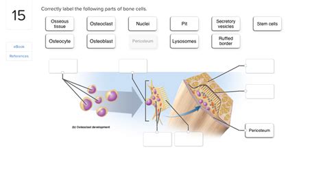
Table of Contents
Correctly Labeling the Parts of Bone Cells: A Comprehensive Guide
Bone, a seemingly inert and static tissue, is in fact a dynamic and highly active organ, constantly undergoing remodeling and renewal. This process is orchestrated by a variety of specialized cells, each with unique structures and functions. Understanding the intricate details of these bone cells, and their constituent parts, is crucial for comprehending bone physiology, pathology, and the development of effective treatments for bone-related diseases. This comprehensive guide will delve into the microscopic anatomy of bone cells, providing a detailed explanation of their key components and their roles in maintaining skeletal health.
The Major Players: Osteoblasts, Osteocytes, and Osteoclasts
Three primary cell types dominate bone tissue: osteoblasts, osteocytes, and osteoclasts. Each possesses distinctive characteristics reflected in their morphology and function. Let's explore the intricacies of each:
Osteoblasts: The Bone Builders
Osteoblasts are the bone-forming cells, responsible for synthesizing and secreting the organic components of the bone matrix, primarily type I collagen and other proteins. These components form the framework upon which mineral deposition occurs, resulting in the hardened, mineralized bone tissue. Here's a breakdown of their key parts:
-
Rough Endoplasmic Reticulum (RER): Highly developed in osteoblasts, the RER is responsible for protein synthesis. Given the significant role of collagen in bone formation, the extensive RER is critical for producing sufficient quantities of this structural protein. Abundant ribosomes stud the RER, highlighting the intense protein synthesis activity within these cells.
-
Golgi Apparatus: This organelle processes and packages proteins synthesized in the RER. In osteoblasts, the Golgi apparatus plays a vital role in preparing the collagen molecules for secretion into the extracellular matrix. It also modifies and packages other proteins crucial for bone formation and mineralization.
-
Secretory Vesicles: These membrane-bound sacs transport the processed collagen and other matrix proteins from the Golgi apparatus to the cell membrane. They then fuse with the membrane, releasing their contents into the extracellular space, where they contribute to the growing bone matrix. The size and number of these vesicles directly reflect the osteoblast's activity level.
-
Alkaline Phosphatase (ALP): This enzyme is a key marker of osteoblast activity. It plays a vital role in the mineralization process, facilitating the deposition of calcium phosphate crystals onto the collagen framework, thus hardening the bone matrix. High ALP levels are indicative of active bone formation.
-
Cell Membrane: The osteoblast's cell membrane regulates the transport of ions and molecules between the cell and its surroundings. It's particularly important for the regulated secretion of the bone matrix components and the uptake of minerals for mineralization. Specific membrane proteins facilitate this selective transport.
-
Nucleus: Contains the genetic material (DNA) that directs all cellular activities, including the synthesis of bone matrix proteins. The nucleus's size and structure can reflect the osteoblast's metabolic activity and stage of differentiation.
Osteocytes: The Bone Maintainers
Osteocytes, derived from osteoblasts, are the most abundant cells in mature bone tissue. They reside within lacunae, small spaces within the mineralized bone matrix, connected to each other and to the bone surface via a network of canaliculi. Their main function is to maintain bone tissue homeostasis, sensing mechanical stress and regulating bone remodeling.
-
Cell Body (within Lacuna): The osteocyte's cell body is located within the lacuna, a cavity in the bone matrix. This location allows for efficient communication and sensing of mechanical forces acting on the bone. The shape of the cell body often reflects the surrounding matrix structure.
-
Canaliculi: These are tiny channels extending from the lacunae, connecting adjacent osteocytes and allowing for communication and exchange of nutrients and waste products. The extensive canalicular network ensures that even osteocytes deep within the bone tissue receive adequate supplies. The density of canaliculi is directly related to the bone's overall health and structural integrity.
-
Gap Junctions: These specialized cell junctions connect neighboring osteocytes within the canaliculi, facilitating direct cell-to-cell communication. Gap junctions allow for the rapid transmission of signals and nutrients between osteocytes, coordinating bone remodeling and responding to mechanical stress. The number and functionality of gap junctions are crucial for the proper functioning of the osteocyte network.
-
Cytoplasmic Processes: These long, slender extensions of the osteocyte's cytoplasm extend into the canaliculi. They are crucial for communication and nutrient exchange between osteocytes and for sensing mechanical stimuli. The length and branching pattern of these processes can vary depending on the osteocyte's location and function.
-
Nucleus: Similar to osteoblasts, the osteocyte nucleus contains the genetic information controlling the cell's functions. However, the osteocyte nucleus is generally smaller and less prominent than that of an osteoblast, reflecting its lower rate of protein synthesis.
Osteoclasts: The Bone Resorbers
Osteoclasts are large, multinucleated cells responsible for bone resorption, the process of breaking down bone tissue. This crucial function is essential for bone remodeling, allowing for the removal of damaged or old bone and the release of calcium and phosphate into the bloodstream.
-
Multinucleated Cytoplasm: Osteoclasts are distinguished by their large size and the presence of multiple nuclei, typically ranging from two to over fifty. The presence of multiple nuclei reflects the cell's high level of metabolic activity. The number of nuclei can be an indicator of the osteoclast's resorptive capacity.
-
Ruffled Border: This highly specialized membrane region is the site of bone resorption. It's characterized by a complex, folded structure that increases the surface area for the secretion of acids and enzymes that dissolve the bone mineral and matrix. The extent of the ruffled border's development is directly related to the osteoclast's activity.
-
Clear Zone: This region surrounds the ruffled border, forming a sealed compartment where bone resorption takes place. This sealing zone isolates the resorption site, preventing the secreted acids and enzymes from damaging surrounding tissues. The integrity of this zone is crucial for the controlled and efficient resorption process.
-
Lysosomes: These organelles contain powerful enzymes capable of breaking down the organic components of the bone matrix, such as collagen. The release of these enzymes into the resorption compartment contributes to the breakdown of the bone matrix. The number and activity of lysosomes reflect the osteoclast's resorptive capacity.
-
Proton Pumps: These specialized proteins in the ruffled border pump hydrogen ions (H+) into the resorption compartment, creating an acidic environment that dissolves the bone mineral. The efficiency of these pumps is directly linked to the rate of bone resorption.
The Interplay of Bone Cells: A Dynamic Equilibrium
These three cell types – osteoblasts, osteocytes, and osteoclasts – work in concert, maintaining a dynamic equilibrium that ensures bone health and structural integrity. This intricate interplay is regulated by various hormonal and local factors, ensuring that bone formation and resorption are appropriately balanced throughout life. Disruptions in this delicate balance can lead to a variety of bone diseases, highlighting the crucial role of each cell type and their constituent parts.
Clinical Significance and Future Directions
Understanding the structure and function of bone cells is not just an academic pursuit; it has significant clinical implications. Conditions such as osteoporosis, Paget's disease, and various bone cancers are all characterized by imbalances in bone remodeling, often involving dysregulation of osteoblast, osteocyte, or osteoclast activity. Detailed knowledge of these cells allows for the development of targeted therapies to address these imbalances and improve bone health.
Future research focusing on bone cell biology holds immense potential for developing innovative treatments. This includes exploring novel drug targets that can selectively modulate the activity of osteoblasts and osteoclasts, potentially leading to more effective treatments for osteoporosis and other bone diseases. Further investigation into the intricate communication networks between bone cells, particularly the role of osteocytes in sensing mechanical stress and regulating bone remodeling, could pave the way for developing strategies to stimulate bone formation and improve bone quality. By continuing to unravel the mysteries of these fascinating cells, we can move closer to a future where bone diseases are effectively prevented and treated.
Conclusion
The precise labeling of the parts of bone cells is crucial for a thorough understanding of bone biology and its clinical implications. From the protein-synthesizing machinery of osteoblasts to the intricate communication network of osteocytes and the bone-resorbing prowess of osteoclasts, each component contributes to the overall health and functionality of the skeletal system. Further research in this field will undoubtedly lead to advancements in the prevention and treatment of bone diseases, improving the quality of life for millions.
Latest Posts
Latest Posts
-
4 05 Quiz Buying Food And Eating Out 1
Mar 19, 2025
-
Unit 6 Test Similar Triangles Answers Pdf
Mar 19, 2025
-
On Psychiatric Units The Most Frequent Victims Of Assault Are
Mar 19, 2025
-
Which Of The Following Is Not Used For Authentication
Mar 19, 2025
-
Upon Your Release A Dod Public Affairs
Mar 19, 2025
Related Post
Thank you for visiting our website which covers about Correctly Label The Following Parts Of Bone Cells . We hope the information provided has been useful to you. Feel free to contact us if you have any questions or need further assistance. See you next time and don't miss to bookmark.
