Correctly Label The Following Tissues Of The Digestive Tract
Onlines
Mar 10, 2025 · 6 min read
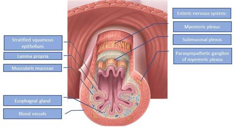
Table of Contents
Correctly Labeling the Tissues of the Digestive Tract: A Comprehensive Guide
The digestive tract, a marvel of biological engineering, is responsible for the breakdown and absorption of nutrients essential for life. Understanding its intricate structure, particularly the diverse tissues that compose its layers, is crucial for comprehending its function and potential pathologies. This comprehensive guide will delve into the detailed histological organization of the digestive tract, focusing on the correct labeling of its key tissues. We'll explore each layer—from the mucosa to the adventitia—and the specific cell types and structures within, providing a firm foundation for anyone studying digestive physiology or pathology.
The Four Basic Layers of the Digestive Tract
The digestive tract, from the esophagus to the anus, exhibits a consistent structural plan, characterized by four primary layers:
- Mucosa: The innermost layer, directly contacting the ingested material. It's responsible for secretion, absorption, and protection.
- Submucosa: A layer of connective tissue rich in blood vessels, lymphatics, and the submucosal plexus (Meissner's plexus), a component of the enteric nervous system.
- Muscularis Externa: Composed of smooth muscle, responsible for peristalsis and segmentation, crucial for the movement of food through the tract. It also contains the myenteric plexus (Auerbach's plexus), another part of the enteric nervous system.
- Adventitia/Serosa: The outermost layer. The esophagus is covered by adventitia (fibrous connective tissue), while the rest of the digestive tract (stomach, small intestine, large intestine) is covered by serosa (a serous membrane composed of a thin layer of connective tissue covered by mesothelium).
Detailed Analysis of Each Layer and its Tissue Components
Let's now examine each layer in detail, highlighting the key tissues and their functions:
1. The Mucosa: A Tripartite Layer
The mucosa is not a single tissue but rather a composite layer with three components:
-
Epithelium: The innermost lining, directly interacting with the lumen. The type of epithelium varies depending on the location within the digestive tract. For example:
- Esophagus: Stratified squamous non-keratinized epithelium, providing protection against abrasion.
- Stomach: Simple columnar epithelium with gastric pits leading to gastric glands, secreting mucus, acid, and digestive enzymes. Specific cell types include mucous neck cells, parietal cells (HCl secretion), chief cells (pepsinogen secretion), and enteroendocrine cells (hormone secretion).
- Small Intestine: Simple columnar epithelium with villi and microvilli, maximizing surface area for absorption. Specialized cells include absorptive enterocytes, goblet cells (mucus secretion), Paneth cells (antimicrobial peptide secretion), and enteroendocrine cells.
- Large Intestine: Simple columnar epithelium with goblet cells predominating, producing mucus for lubrication and protection.
-
Lamina Propria: A layer of loose connective tissue underlying the epithelium. It contains blood vessels, lymphatics, immune cells (e.g., lymphocytes, plasma cells), and nerve fibers. In the small intestine, the lamina propria extends into the villi, enhancing absorptive capacity.
-
Muscularis Mucosae: A thin layer of smooth muscle, responsible for local movements of the mucosa, enhancing secretion and absorption.
2. The Submucosa: A Support Layer
The submucosa is a layer of dense irregular connective tissue that provides structural support to the mucosa. Its key features include:
- Blood Vessels and Lymphatics: A rich network supplying the mucosa and carrying away absorbed nutrients.
- Submucosal Plexus (Meissner's Plexus): A component of the enteric nervous system, regulating local blood flow, secretion, and absorption.
- Meissner's Plexus: This network of neurons coordinates the activity of the mucosa, controlling secretions and local blood flow. It's crucial for maintaining the mucosal environment optimal for digestion and absorption.
3. The Muscularis Externa: The Engine of Movement
The muscularis externa is responsible for the propulsion of food along the digestive tract through peristalsis and segmentation. Its structure varies depending on location:
-
Esophagus: Contains both skeletal and smooth muscle. The upper third is predominantly skeletal muscle (under voluntary control), while the lower two-thirds transition to smooth muscle (involuntary control).
-
Stomach: Has three layers: an inner oblique layer, a middle circular layer, and an outer longitudinal layer. This complex arrangement allows for churning and mixing of food.
-
Small and Large Intestines: Typically consist of two layers: an inner circular layer and an outer longitudinal layer. The longitudinal layer in the large intestine is thickened into three bands called teniae coli.
-
Myenteric Plexus (Auerbach's Plexus): Located between the circular and longitudinal muscle layers, this is another key part of the enteric nervous system, regulating the motility of the digestive tract. It coordinates peristalsis and segmentation.
4. The Adventitia/Serosa: The Outermost Covering
The outermost layer varies depending on the location within the digestive tract:
- Adventitia: Found in the esophagus, it's composed of loose connective tissue that blends with the surrounding tissues of the mediastinum. It provides support and anchors the esophagus.
- Serosa: Covering the stomach, small intestine, and large intestine, it’s a serous membrane composed of a thin layer of loose connective tissue covered by a simple squamous mesothelium. The serosa reduces friction between the digestive organs and the surrounding abdominal cavity. The serosa is part of the peritoneum.
Variations in Tissue Composition Along the Digestive Tract
It's crucial to understand that the specific tissue composition within each layer varies significantly along the length of the digestive tract, reflecting the diverse functions of different regions.
- The esophagus prioritizes protection against abrasion with its stratified squamous epithelium.
- The stomach is specialized for secretion of acid and digestive enzymes with its gastric glands and specific cell types.
- The small intestine excels at absorption with its villi and microvilli, abundant capillaries, and specialized absorptive cells.
- The large intestine focuses on water absorption and waste elimination with its abundance of goblet cells and simpler epithelium.
Understanding these variations is critical for correctly interpreting histological images and comprehending the physiological processes occurring in different parts of the digestive system.
Clinical Significance of Digestive Tract Histology
Incorrect labeling of tissues can lead to misdiagnosis and inappropriate treatment. Accurate identification of the tissues of the digestive tract is essential in several clinical contexts:
- Diagnosis of Inflammatory Bowel Disease (IBD): Histological examination of biopsies is crucial for distinguishing between Crohn's disease and ulcerative colitis. Specific inflammatory changes in the mucosa and submucosa are characteristic of each condition.
- Detection of Gastric Cancer: Histological analysis of tissue samples allows for the identification of cancerous cells and staging of the tumor, guiding treatment decisions.
- Assessment of Celiac Disease: Biopsy of the small intestine reveals characteristic villous atrophy and inflammation in individuals with celiac disease.
- Identification of Infections: Histological examination can help identify pathogens such as Helicobacter pylori (associated with gastric ulcers) or parasitic infections.
Precise identification of all tissue layers and constituent cells is paramount for accurate diagnosis and effective management of various gastrointestinal disorders. Proper labeling of tissue samples is, therefore, not just an academic exercise but a crucial step in the diagnosis and treatment of gastrointestinal diseases.
Conclusion
Correctly labeling the tissues of the digestive tract requires a detailed understanding of the four basic layers—mucosa, submucosa, muscularis externa, and adventitia/serosa—and the specific tissues and cell types within each layer. The variation in tissue composition along the tract reflects the specialized functions of different regions. This knowledge is not only essential for academic understanding but also plays a crucial role in clinical diagnosis and treatment of a wide range of gastrointestinal disorders. By mastering the intricate histology of the digestive tract, we gain a deeper appreciation for the complexity and elegance of this vital system. Continued study and meticulous attention to detail are vital for accurately identifying and labeling these tissues.
Latest Posts
Latest Posts
-
Apush Unit 3 Progress Check Mcq
Mar 10, 2025
-
What Is Brand Association Select All That Apply
Mar 10, 2025
-
Basic Communication Crossword Notes Puzzle Answers
Mar 10, 2025
-
How Do You Individualize A Patients Care Plan In Epic
Mar 10, 2025
-
Rising Action Examples In Helen Keller
Mar 10, 2025
Related Post
Thank you for visiting our website which covers about Correctly Label The Following Tissues Of The Digestive Tract . We hope the information provided has been useful to you. Feel free to contact us if you have any questions or need further assistance. See you next time and don't miss to bookmark.
