Correctly Label The Muscles Of The Leg
Onlines
Mar 10, 2025 · 7 min read
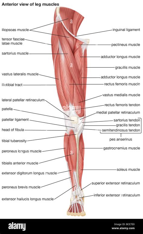
Table of Contents
Correctly Label the Muscles of the Leg: A Comprehensive Guide
Knowing the muscles of the leg is crucial for anyone involved in fitness, physical therapy, sports medicine, or simply understanding the human body. This comprehensive guide will help you correctly label the muscles of the leg, breaking them down into compartments and providing detailed descriptions to aid in memorization and understanding. We'll cover the anterior, posterior, and lateral compartments of both the thigh and leg, highlighting key functions and clinical considerations.
Understanding Leg Muscle Organization
The muscles of the leg are organized into compartments, separated by strong fascia (connective tissue). This compartmentalization is essential for understanding muscle function and clinical presentations of injuries. Understanding the leg's anatomy involves knowing the different compartments and their associated muscles. Let’s dive into each compartment.
The Thigh: A Deep Dive into Compartments
The thigh, the segment of the lower limb between the hip and the knee, contains three main compartments: anterior, medial, and posterior.
1. Anterior Compartment of the Thigh
The anterior compartment of the thigh is primarily responsible for knee extension and hip flexion. The key players here are:
-
Quadriceps Femoris: This is actually a group of four muscles, collectively responsible for powerful knee extension. They are:
- Rectus Femoris: Unique among the quadriceps as it's a two-joint muscle, contributing to both hip flexion and knee extension. It originates from the anterior inferior iliac spine and the superior acetabulum of the hip bone.
- Vastus Lateralis: The largest of the quadriceps, situated on the outer side of the thigh. Its primary action is knee extension.
- Vastus Medialis: Located on the inner side of the thigh, contributing significantly to knee extension and medial patellar stabilization.
- Vastus Intermedius: Lies deep to the rectus femoris, contributing to knee extension. It's often difficult to palpate.
-
Sartorius: This long, thin muscle is the longest muscle in the body. It acts as a weak hip flexor, abductor, and lateral rotator, and also assists in knee flexion.
Clinical Significance: Quadriceps injuries are common in athletic populations, ranging from strains to tears. Patellofemoral pain syndrome (runner's knee) often involves dysfunction within the quadriceps and patellar tracking.
2. Medial Compartment of the Thigh
The medial compartment of the thigh, also known as the adductor compartment, primarily focuses on hip adduction. The key muscles include:
- Adductor Longus: A superficial muscle contributing to hip adduction, flexion, and medial rotation.
- Adductor Brevis: A deeper muscle situated beneath the adductor longus, also performing hip adduction.
- Adductor Magnus: The largest of the adductor muscles, with a complex structure and diverse functions including hip adduction, extension, and medial rotation. It has two distinct heads: the adductor part and the hamstring part.
- Gracilis: A thin muscle located medially on the thigh, contributing to hip adduction and knee flexion.
Clinical Significance: Adductor strains are common in athletes, particularly in sports involving rapid changes in direction. Adductor longus tendinopathy can also cause pain in the groin area.
3. Posterior Compartment of the Thigh (Hamstrings)
The posterior compartment of the thigh houses the hamstring muscles, primarily responsible for hip extension and knee flexion. The hamstrings are crucial for many movements and activities. They include:
- Biceps Femoris: Located on the lateral aspect of the thigh, it has two heads (long and short) and performs hip extension and knee flexion and external rotation.
- Semitendinosus: A long, thin muscle located medially, contributing to hip extension and knee flexion and internal rotation.
- Semimembranosus: A broad, flat muscle located medially, deep to the semitendinosus. It also contributes to hip extension and knee flexion and internal rotation.
Clinical Significance: Hamstring strains are among the most frequent muscle injuries in athletes. Overstretching or forceful contractions can lead to varying degrees of muscle damage.
The Leg: Compartmentalization Below the Knee
Below the knee, the leg is divided into three compartments: anterior, lateral, and posterior.
1. Anterior Compartment of the Leg
The anterior compartment of the leg is primarily responsible for dorsiflexion (lifting the foot towards the shin) and toe extension. Its key muscles include:
- Tibialis Anterior: This muscle is essential for dorsiflexion and inversion (turning the sole of the foot inwards).
- Extensor Hallucis Longus: This extends the big toe and contributes to dorsiflexion.
- Extensor Digitorum Longus: This extends the toes (excluding the big toe) and contributes to dorsiflexion.
- Peroneus Tertius: This muscle assists in dorsiflexion and eversion (turning the sole of the foot outwards).
Clinical Significance: Anterior compartment syndrome is a serious condition characterized by increased pressure within the anterior compartment, potentially compromising blood supply to the muscles and nerves.
2. Lateral Compartment of the Leg
The lateral compartment of the leg primarily focuses on eversion (turning the sole of the foot outwards) and assists with plantarflexion (pointing the toes downwards). Its muscles are:
- Peroneus Longus: This muscle contributes to eversion and plantarflexion. It has a unique course, passing behind the lateral malleolus and under the foot.
- Peroneus Brevis: This muscle is primarily involved in eversion and plantarflexion.
Clinical Significance: Peroneal tendon injuries are common, often occurring during strenuous activities or ankle sprains.
3. Posterior Compartment of the Leg (Superficial and Deep)
The posterior compartment of the leg, like the thigh's posterior compartment, is complex and is further subdivided into superficial and deep groups. The muscles in this compartment primarily contribute to plantarflexion, toe flexion, and inversion.
-
Superficial Group:
- Gastrocnemius: A large, superficial muscle forming the bulk of the calf. It contributes to plantarflexion and knee flexion.
- Soleus: Lies deep to the gastrocnemius, playing a key role in plantarflexion.
- Plantaris: A small muscle with a relatively minor role in plantarflexion, often absent in some individuals.
-
Deep Group:
- Tibialis Posterior: This muscle plays a crucial role in plantarflexion and inversion.
- Flexor Hallucis Longus: Flexes the big toe and contributes to plantarflexion and inversion.
- Flexor Digitorum Longus: Flexes the toes (excluding the big toe) and contributes to plantarflexion and inversion.
Clinical Significance: Gastrocnemius and soleus strains are common, often resulting from overuse or sudden forceful contractions. Achilles tendinitis, an inflammation of the Achilles tendon, affects the insertion of the gastrocnemius and soleus muscles. Tibial posterior tendinopathy is also a frequent occurrence, especially in those with flat feet.
Practical Tips for Correctly Labeling Leg Muscles
Learning to label the muscles of the leg requires consistent effort and a multifaceted approach. Here are some practical tips:
- Visual Learning: Use anatomical charts, atlases, and 3D models. Regularly reviewing these visuals will significantly enhance memorization.
- Hands-on Learning: If possible, palpate the muscles on yourself or a partner. Feel the muscle’s shape, texture, and location while referring to anatomical diagrams.
- Mnemonic Devices: Create memory aids using acronyms, rhymes, or stories to help recall the muscle names and their functions.
- Flashcards: Make flashcards with the muscle name on one side and its location, function, and origin/insertion on the other.
- Quizzes and Tests: Regularly test yourself to assess your progress and identify areas needing improvement.
- Clinical Correlation: Relate the muscle actions to everyday movements and activities. Understanding the function will assist in memorization.
- Progressive Learning: Don't try to learn everything at once. Focus on one compartment at a time, mastering it before moving to the next.
Conclusion: Mastering Leg Muscle Anatomy
Correctly labeling the muscles of the leg is a journey that requires dedication and a multi-sensory approach. By combining visual learning, hands-on experience, mnemonic devices, and regular self-testing, you can achieve mastery of this complex but fascinating aspect of human anatomy. This knowledge is valuable not only for academic pursuits but also for those working in healthcare, fitness, or sports-related fields. Remember to continually review and reinforce your learning to ensure long-term retention. The detailed understanding presented here provides a solid foundation for your journey towards complete mastery of leg muscle anatomy.
Latest Posts
Latest Posts
-
Grade 10 Fsa Ela Reading Practice Test Answer Key
Mar 10, 2025
-
The Man To Send Rain Clouds Summary
Mar 10, 2025
-
Excel 365 2021 Capstone Level 1 Working With Sales Data
Mar 10, 2025
-
Fundamentals Of Electric Circuits 7th Edition Solutions Pdf
Mar 10, 2025
-
It Is Always Best To Avoid Conducting Nonexperimental Research
Mar 10, 2025
Related Post
Thank you for visiting our website which covers about Correctly Label The Muscles Of The Leg . We hope the information provided has been useful to you. Feel free to contact us if you have any questions or need further assistance. See you next time and don't miss to bookmark.
