Correctly Label The Parts Of An Exocrine Gland
Onlines
Mar 19, 2025 · 6 min read
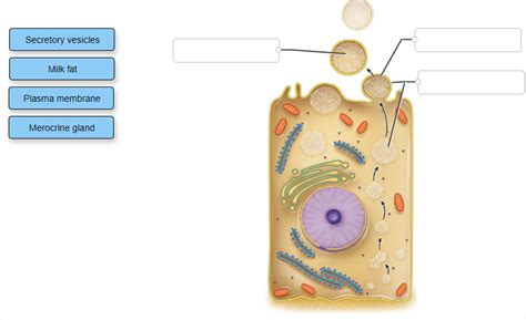
Table of Contents
- Correctly Label The Parts Of An Exocrine Gland
- Table of Contents
- Correctly Labeling the Parts of an Exocrine Gland: A Comprehensive Guide
- The Fundamental Components of Exocrine Glands
- 1. Secretory Unit (Acini/Alveoli):
- 2. Ducts:
- 3. Myoepithelial Cells:
- 4. Connective Tissue Stroma:
- Classifying Exocrine Glands: A Structural Approach
- 1. Based on the structure of the secretory unit:
- 2. Based on the mode of secretion:
- Detailed Examples: Labeling Specific Exocrine Glands
- 1. Salivary Gland (Merocrine):
- 2. Sebaceous Gland (Holocrine):
- 3. Mammary Gland (Apocrine):
- Beyond the Basics: Advanced Aspects of Exocrine Gland Anatomy
- Conclusion: Mastering the Art of Exocrine Gland Labeling
- Latest Posts
- Latest Posts
- Related Post
Correctly Labeling the Parts of an Exocrine Gland: A Comprehensive Guide
Exocrine glands, unlike their endocrine counterparts, secrete their products onto epithelial surfaces, rather than directly into the bloodstream. Understanding their intricate structure is crucial for comprehending their diverse functions throughout the body. This comprehensive guide will delve into the detailed anatomy of exocrine glands, focusing on correctly identifying and labeling their key components. We will explore various types of exocrine glands, highlighting their structural variations and functional implications. By the end, you will possess a thorough understanding of exocrine gland anatomy, enabling accurate labeling and a deeper appreciation of their physiological roles.
The Fundamental Components of Exocrine Glands
All exocrine glands, regardless of their specific type or location, share certain fundamental structural elements. Mastering these foundational components is the first step towards accurate labeling. These key structures include:
1. Secretory Unit (Acini/Alveoli):
This is the functional unit of the exocrine gland, responsible for synthesizing and secreting the gland's product. The secretory unit can take several forms, influencing the classification of the exocrine gland itself. These forms include:
- Acinar/Alveolar: These secretory units are rounded or sac-like. They often appear as clusters of grape-like structures.
- Tubular: These secretory units are elongated and tube-shaped.
- Tubuloacinar/Tubuloalveolar: These glands possess a combination of tubular and acinar/alveolar secretory units.
Accurate labeling: When labeling a diagram, clearly identify the secretory unit type (acinar, tubular, or tubuloacinar) and indicate the location of the secretory cells within the unit.
2. Ducts:
The ducts are a network of tubes that transport the secreted product from the secretory unit to the epithelial surface. The complexity of the duct system varies depending on the gland's size and type. Larger glands often have a more elaborate branched duct system, whereas smaller glands may have simpler, unbranched ducts. The ducts are lined with epithelial cells that can vary in structure and function, depending on their location within the duct system.
Accurate labeling: When labeling, differentiate between the intralobular ducts (within a lobule) and the interlobular ducts (between lobules). Also, note any differences in epithelial cell types lining different parts of the duct system.
3. Myoepithelial Cells:
These specialized cells are located between the secretory cells and the basement membrane. They possess contractile properties, playing a crucial role in propelling the secreted product along the ducts. Their contractile activity is regulated by neural and hormonal signals.
Accurate labeling: These cells are often overlooked, but their inclusion is important for a complete and accurate depiction of exocrine gland structure. Label them clearly and indicate their location relative to the secretory cells and the basement membrane.
4. Connective Tissue Stroma:
Exocrine glands are embedded within a supporting framework of connective tissue. This stroma provides structural support, supplies blood vessels and nerves, and separates different glandular units (lobules). The connective tissue also contains immune cells and contributes to the overall organization of the gland.
Accurate labeling: Identify the connective tissue stroma and highlight its role in supporting and organizing the gland's structure. You can differentiate between the denser connective tissue surrounding the gland (capsule) and the more delicate stroma within the gland.
Classifying Exocrine Glands: A Structural Approach
Exocrine glands are classified based on several criteria, predominantly their secretory unit morphology and the mode of secretion. These classifications are key to understanding the functional diversity of exocrine glands and crucial for accurate labeling.
1. Based on the structure of the secretory unit:
- Simple Glands: Possess a single, unbranched duct.
- Compound Glands: Have a branched duct system.
Within these broad categories, further classification is based on the shape of the secretory unit (acinar, tubular, or tubuloacinar). Therefore, you could have a simple acinar gland, a compound tubular gland, or a compound tubuloacinar gland, each with distinct structural features.
2. Based on the mode of secretion:
- Merocrine Secretion: The secretory product is released via exocytosis, without any damage to the secretory cell. This is the most common mode of secretion. Examples include salivary glands and sweat glands.
- Apocrine Secretion: A portion of the apical cytoplasm is released along with the secretory product. This process involves some degree of cellular damage, which is subsequently repaired. The mammary glands are a prime example.
- Holocrine Secretion: The entire secretory cell undergoes lysis and is released as the secretory product. This is a destructive process, with the secretory cells continuously being replaced. Sebaceous glands exemplify this type of secretion.
Accurate labeling: When labeling, clearly indicate the mode of secretion (merocrine, apocrine, or holocrine) and any visible structural features associated with that mode. For example, in holocrine glands, you might label cellular debris within the lumen of the secretory unit.
Detailed Examples: Labeling Specific Exocrine Glands
Let’s apply our knowledge to label the specific parts of some common exocrine glands.
1. Salivary Gland (Merocrine):
A salivary gland is a classic example of a compound tubuloacinar gland. When labeling a diagram, ensure you clearly identify:
- Serous acini: These produce a watery secretion rich in enzymes.
- Mucous acini: These produce a viscous, mucus-like secretion.
- Intercalated ducts: Small ducts that drain the acini.
- Intralobular ducts: Ducts within a lobule.
- Interlobular ducts: Larger ducts between lobules.
- Main excretory duct: The large duct that carries saliva to the oral cavity.
- Myoepithelial cells: Located between the secretory cells and the basement membrane.
- Connective tissue stroma: The supporting connective tissue.
2. Sebaceous Gland (Holocrine):
Sebaceous glands are simple alveolar glands with a holocrine mode of secretion. Key features to label:
- Secretory acini: Filled with lipid-rich secretory product and cellular debris.
- Short duct: Drains into a hair follicle or directly onto the skin surface.
- Connective tissue capsule: Surrounds the gland.
The absence of distinct intercalated and intralobular ducts, as seen in salivary glands, is a crucial differentiating factor.
3. Mammary Gland (Apocrine):
Mammary glands are complex tubuloalveolar glands with an apocrine mode of secretion. Labeling should include:
- Alveoli: Milk-producing units.
- Ducts: A branched network of ducts draining the alveoli.
- Lactiferous sinuses: Larger ducts near the nipple.
- Myoepithelial cells: Surrounding the alveoli and ducts.
- Connective tissue stroma: Support structure.
The presence of the alveoli, responsible for milk production, and the more complex duct system distinguishes the mammary gland from other exocrine glands.
Beyond the Basics: Advanced Aspects of Exocrine Gland Anatomy
While the core components discussed above provide a solid foundation, there are several more nuanced aspects worth exploring for a truly comprehensive understanding.
- Innervation: The intricate nerve supply regulating glandular activity, including secretion and ductal contraction.
- Vascular Supply: The rich network of blood vessels providing nutrients and removing waste products.
- Cellular heterogeneity: The presence of various cell types within the gland, beyond the secretory and ductal cells. This includes immune cells and other supporting cells.
- Developmental Aspects: Tracing the developmental origins and differentiation pathways of exocrine glands.
- Pathophysiology: Exploring the various diseases and conditions affecting exocrine glands.
Including these advanced aspects in your labeling and understanding will elevate your knowledge beyond the basic framework.
Conclusion: Mastering the Art of Exocrine Gland Labeling
Correctly labeling the parts of an exocrine gland is a crucial skill for anyone studying anatomy, histology, or physiology. By understanding the fundamental components—the secretory unit, ducts, myoepithelial cells, and connective tissue stroma—and the various classification systems, you can accurately and comprehensively represent the complex structure and function of these vital glands. Remember to consider the specific features of each gland type, their mode of secretion, and the nuances of their internal organization. With practice and attention to detail, you will master the art of exocrine gland labeling and gain a deeper appreciation for their important role in maintaining bodily homeostasis.
Latest Posts
Latest Posts
-
Under Dodd 5240 06 Reportable Foreign Intelligence
Mar 20, 2025
-
What Sata Devices Did You Find
Mar 20, 2025
-
End Of Semester Test English 11a
Mar 20, 2025
-
Where Is An Integrated Microphone Normally Located On A Laptop
Mar 20, 2025
-
Pogil Electron Configuration And Orbitals Answer Key Model 2
Mar 20, 2025
Related Post
Thank you for visiting our website which covers about Correctly Label The Parts Of An Exocrine Gland . We hope the information provided has been useful to you. Feel free to contact us if you have any questions or need further assistance. See you next time and don't miss to bookmark.
