Correctly Label The Pectoral And Brachial Muscles
Onlines
Mar 31, 2025 · 6 min read
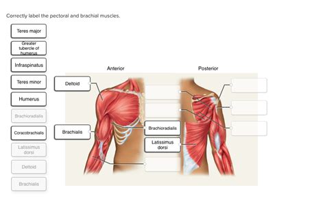
Table of Contents
Correctly Labeling the Pectoral and Brachial Muscles: A Comprehensive Guide
Understanding the intricate network of muscles in the chest and upper arm is crucial for anyone involved in anatomy, fitness, physical therapy, or sports medicine. This comprehensive guide will delve deep into the pectoral and brachial muscle groups, providing detailed descriptions, origins, insertions, actions, and innervations to ensure accurate labeling. We'll also explore common misconceptions and provide practical tips for mastering this complex anatomical region.
The Pectoral Muscles: Powerhouses of the Chest
The pectoral muscles, located on the anterior (front) of the chest, play a vital role in arm movement, shoulder stability, and overall upper body strength. They are broadly categorized into two main groups: the pectoralis major and the pectoralis minor.
Pectoralis Major: The "Big Chest" Muscle
The pectoralis major is a large, fan-shaped muscle dominating the anterior chest. Its prominent size and superficial location make it easily identifiable. It consists of three distinct parts:
- Clavicular Head: Originating from the medial half of the clavicle (collarbone).
- Sternocostal Head: Originating from the sternum (breastbone) and the upper six costal cartilages (cartilages connecting the ribs to the sternum).
- Abdominal Head: Originating from the aponeurosis of the external oblique muscle (abdominal muscle).
Insertion: All three heads converge to insert into the greater tubercle of the humerus (upper arm bone).
Action: The pectoralis major performs a variety of actions, depending on the part engaged and the position of the arm:
- Adduction: Bringing the arm closer to the body.
- Internal Rotation: Rotating the arm inward towards the body.
- Horizontal Adduction: Bringing the arms across the body.
- Flexion: Raising the arm forward (primarily the clavicular head).
Innervation: The pectoralis major is innervated by the medial and lateral pectoral nerves (C5-T1).
Clinical Significance: Injuries to the pectoralis major, often involving tears or strains, are common in athletes, particularly those involved in weightlifting or throwing sports. These injuries often present with pain, weakness, and deformity in the chest region.
Pectoralis Minor: The Deeper Chest Muscle
Lying deep beneath the pectoralis major, the pectoralis minor is a smaller, triangular muscle.
Origin: The third to fifth ribs.
Insertion: Coracoid process of the scapula (shoulder blade).
Action: The pectoralis minor primarily assists in protraction (moving the scapula forward) and downward rotation of the scapula. It also contributes to rib cage elevation during forced inspiration.
Innervation: Medial pectoral nerve (C8-T1).
Clinical Significance: While less prone to injury than the pectoralis major, the pectoralis minor can be involved in conditions such as thoracic outlet syndrome, where compression of nerves and blood vessels in the space between the clavicle and first rib causes pain and numbness in the arm and hand.
The Brachial Muscles: Movers of the Arm
The brachial muscles are located in the upper arm and are responsible for a wide range of arm movements, including flexion, extension, and supination (turning the palm upward). These muscles are generally divided into anterior (front) and posterior (back) compartments.
Anterior Compartment of the Brachium: Flexors
This compartment primarily houses muscles responsible for elbow flexion (bending the elbow) and forearm supination.
-
Biceps Brachii: The most superficial muscle in the anterior compartment, with two heads:
- Long Head: Originates from the supraglenoid tubercle of the scapula.
- Short Head: Originates from the coracoid process of the scapula.
Insertion: Radial tuberosity of the radius (forearm bone) and bicipital aponeurosis (a fibrous expansion that inserts into the deep fascia of the forearm).
Action: Elbow flexion, forearm supination, and shoulder flexion (long head only).
Innervation: Musculocutaneous nerve (C5-C7).
-
Brachialis: Deep to the biceps brachii, the brachialis is a powerful elbow flexor.
Origin: Distal half of the anterior humerus.
Insertion: Ulnar tuberosity of the ulna (forearm bone).
Action: Elbow flexion.
Innervation: Musculocutaneous nerve (C5-C7).
-
Coracobrachialis: A smaller muscle located medially, assisting in arm flexion and adduction.
Origin: Coracoid process of the scapula.
Insertion: Medial aspect of the humerus.
Action: Shoulder flexion and adduction; weak elbow flexion.
Innervation: Musculocutaneous nerve (C5-C7).
Posterior Compartment of the Brachium: Extensors
This compartment houses muscles responsible for elbow extension (straightening the elbow) and forearm pronation (turning the palm downward).
-
Triceps Brachii: The dominant muscle of the posterior compartment, consisting of three heads:
- Long Head: Originates from the infraglenoid tubercle of the scapula.
- Lateral Head: Originates from the posterior humerus, superior to the radial groove.
- Medial Head: Originates from the posterior humerus, inferior to the radial groove.
Insertion: Olecranon process of the ulna.
Action: Elbow extension, shoulder extension (long head only).
Innervation: Radial nerve (C6-C8).
-
Anconeus: A small muscle assisting in elbow extension and stabilizing the elbow joint.
Origin: Lateral epicondyle of the humerus.
Insertion: Olecranon process of the ulna.
Action: Elbow extension; assists in forearm pronation and supination.
Innervation: Radial nerve (C7-C8).
Common Misconceptions and Labeling Challenges
Accurate labeling of these muscles requires careful attention to detail. Several common misconceptions can lead to errors:
- Confusing Biceps and Triceps Heads: Differentiating the long, short, medial, and lateral heads of the biceps and triceps can be challenging. Pay close attention to their origins and insertions.
- Overlapping Muscles: The superficial location of some muscles can obscure underlying structures. Consider using anatomical charts and models to visualize the layers.
- Variable Muscle Attachments: Slight variations in muscle attachment points can exist between individuals. Understanding the typical ranges is crucial.
Tips for Accurate Labeling
Mastering the labeling of pectoral and brachial muscles requires consistent practice and a multi-faceted approach:
- Utilize Anatomical Resources: Refer to detailed anatomical atlases, textbooks, and online resources. High-quality anatomical illustrations are invaluable.
- Study from Multiple Angles: Examine images and models from different viewpoints – anterior, posterior, lateral – to appreciate the spatial relationships between muscles.
- Palpate the Muscles: Whenever possible, palpate (feel) the muscles on yourself or a partner to gain a kinesthetic understanding of their location and size.
- Relate Function to Form: Understanding the actions of each muscle can help you infer its location and orientation.
- Practice, Practice, Practice: Regular labeling exercises are essential for solidifying your knowledge. Use flashcards, quizzes, and anatomical drawing practice.
Conclusion: Mastering the Anatomy of Movement
Accurate labeling of the pectoral and brachial muscles is fundamental to understanding human movement and function. By utilizing a combination of visual learning, tactile exploration, and consistent practice, you can achieve a thorough grasp of this complex anatomical region. This knowledge is invaluable for professionals in various fields, from fitness instructors and physical therapists to medical students and researchers. Remember that continuous learning and review are essential for maintaining proficiency in anatomical terminology and understanding. The more you study and practice, the easier it will become to correctly identify and label these important muscle groups.
Latest Posts
Latest Posts
-
Speechmaking Loses Value If The Speaker Lacks
Apr 01, 2025
-
What Did The Ape Think Of The Grape House
Apr 01, 2025
-
Unlike Tort Law Contract Law States That
Apr 01, 2025
-
If A Generalization Must Be Made It Should
Apr 01, 2025
-
Which Of These Statements About The Elderly Is True
Apr 01, 2025
Related Post
Thank you for visiting our website which covers about Correctly Label The Pectoral And Brachial Muscles . We hope the information provided has been useful to you. Feel free to contact us if you have any questions or need further assistance. See you next time and don't miss to bookmark.
