Deep Vein Thrombosis Hesi Case Study
Onlines
Mar 14, 2025 · 6 min read
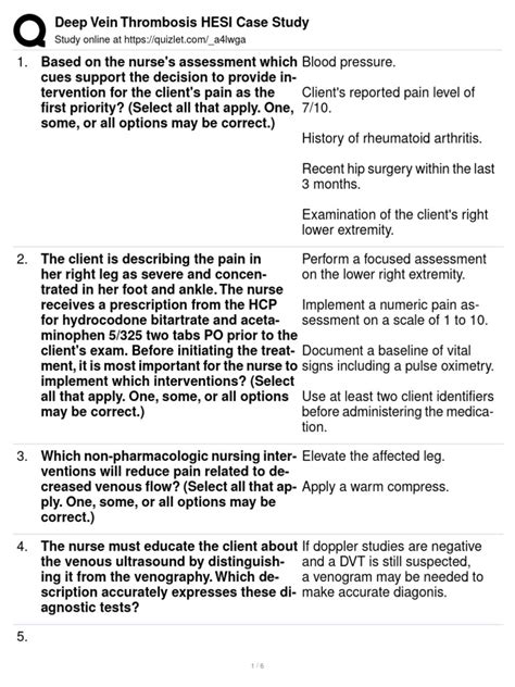
Table of Contents
Deep Vein Thrombosis (DVT): A Comprehensive HESI Case Study Analysis
Deep vein thrombosis (DVT) is a serious medical condition characterized by the formation of a blood clot (thrombus) within a deep vein, most commonly in the legs. This case study will delve into a hypothetical HESI scenario involving a patient with DVT, exploring the pathophysiology, risk factors, clinical manifestations, diagnostic procedures, treatment strategies, and nursing implications. Understanding DVT requires a multifaceted approach encompassing the patient's history, physical assessment findings, and appropriate interventions. This analysis aims to provide a thorough understanding of DVT management within a healthcare setting.
Understanding the Pathophysiology of DVT
DVT arises from a complex interplay of factors known as Virchow's triad:
-
Endothelial damage: Injury to the inner lining of the vein, often due to trauma, surgery, or inflammation, disrupts the smooth flow of blood and promotes clot formation.
-
Venous stasis: Slow or stagnant blood flow allows clotting factors to accumulate, increasing the likelihood of thrombus formation. This is commonly seen in prolonged immobility, such as after surgery or during long periods of sitting or lying down.
-
Hypercoagulability: Conditions that increase blood clotting tendency, such as genetic predisposition, pregnancy, certain medications (oral contraceptives), cancer, and dehydration, significantly elevate DVT risk.
The formation of a thrombus can partially or completely obstruct blood flow, leading to various complications. The clot itself may embolize, meaning a fragment breaks off and travels to the lungs, causing a life-threatening pulmonary embolism (PE).
HESI Case Study Scenario: Presenting Symptoms and Initial Assessment
Patient: 65-year-old female, post-operative day 3 following total knee replacement.
Presenting Symptoms:
- Right leg pain and swelling.
- Localized warmth and erythema (redness) in the right calf.
- Positive Homan's sign (though this is becoming less reliable in diagnosis).
- Mild shortness of breath and chest pain.
Initial Assessment:
Upon initial assessment, the nurse notes the patient's vital signs, including elevated heart rate and respiratory rate, suggesting potential respiratory distress associated with a possible PE. The nurse should also assess the patient's pain level using a validated pain scale (e.g., numeric rating scale). A thorough neurovascular assessment of the affected leg is crucial, including checking for pulses, skin temperature, capillary refill, and sensation. The patient's medical history, including any known clotting disorders, use of medications, and family history of DVT or PE, should be carefully reviewed. Any other symptoms, like unexplained fatigue or fever, should be documented.
Diagnostic Procedures for DVT
Several diagnostic tests are crucial in confirming the diagnosis of DVT:
-
Doppler ultrasound: This non-invasive test uses sound waves to visualize blood flow in the veins. It can detect the presence of a thrombus and assess the extent of venous occlusion. This is usually the first-line diagnostic test for suspected DVT.
-
D-dimer test: This blood test measures the level of D-dimer, a protein fragment released during clot breakdown. Elevated D-dimer levels suggest the presence of a clot, but it's not specific to DVT and can be elevated in other conditions. A negative D-dimer test can effectively rule out DVT.
-
Venography: This invasive procedure involves injecting contrast dye into the veins and using X-rays to visualize the blood vessels. It’s less commonly used now due to the availability of less invasive options.
-
Computed tomography pulmonary angiography (CTPA): This imaging test is used to rule out or confirm a pulmonary embolism (PE), a life-threatening complication of DVT.
Treatment Strategies for DVT
Treatment for DVT aims to prevent clot propagation, reduce the risk of PE, and alleviate symptoms. The primary treatment modalities include:
-
Anticoagulation therapy: This involves administering medications to prevent further clot formation and reduce the risk of embolization. The most commonly used anticoagulants include:
- Heparin: This is usually given intravenously initially to achieve rapid anticoagulation. Low molecular weight heparin (LMWH), such as enoxaparin, is often preferred due to its ease of administration and fewer monitoring requirements.
- Warfarin (Coumadin): This oral anticoagulant is used for long-term prophylaxis, typically after the initial heparin treatment. Regular monitoring of the international normalized ratio (INR) is necessary to ensure therapeutic anticoagulation.
- Direct thrombin inhibitors (e.g., dabigatran): These newer anticoagulants offer convenience and may reduce bleeding risk in some patients. However, they have limitations and specific usage guidelines.
- Direct factor Xa inhibitors (e.g., rivaroxaban, apixaban): Similar to direct thrombin inhibitors, they are a popular alternative to warfarin due to their convenience and reduced need for monitoring.
-
Thrombolytic therapy: This aggressive treatment involves using medications to break down existing clots. It is usually reserved for patients with massive DVT or those at high risk of PE, and it carries a significant risk of bleeding.
-
Inferior vena cava (IVC) filter: In cases where anticoagulation therapy is contraindicated or ineffective, an IVC filter can be placed to prevent emboli from traveling to the lungs. This device acts as a barrier, trapping clots before they can reach the pulmonary circulation.
Nursing Implications in DVT Management
Nursing care for patients with DVT is crucial in preventing complications and promoting optimal outcomes. Key nursing interventions include:
-
Assessment: Continuous monitoring of vital signs, leg circumference, pain levels, and neurovascular status is essential. Early detection of any changes can help prevent serious complications.
-
Pain management: Administering analgesics as prescribed and implementing non-pharmacological pain relief strategies, such as elevation of the affected leg, can significantly improve patient comfort.
-
Medication administration: Administering anticoagulants as ordered and carefully monitoring for adverse effects, such as bleeding, is paramount. Patient education regarding medication adherence and potential side effects is crucial.
-
Mobility and ambulation: As soon as clinically appropriate, encouraging early ambulation and leg exercises helps prevent venous stasis. This should be guided by the patient’s individual condition and physician’s orders. However, excessive activity should be avoided early in the treatment process.
-
Leg elevation: Elevating the affected leg reduces swelling and improves venous return. Patient education regarding proper leg elevation techniques should be provided.
-
Compression therapy: Applying graduated compression stockings can reduce edema and improve venous return, although specific timing regarding their application might vary based on individual cases.
-
Patient education: Educating patients about DVT risk factors, preventive measures, and the importance of adhering to the prescribed treatment plan is crucial for improving long-term outcomes. Patients should be taught to recognize signs and symptoms of worsening DVT or PE and seek immediate medical attention if necessary.
Prevention of DVT
Preventing DVT is crucial, especially in high-risk individuals. Preventive measures include:
-
Early ambulation: Post-operative mobilization, as tolerated, is essential to prevent venous stasis.
-
Leg exercises: Regular ankle pumps, foot circles, and knee flexion exercises improve venous return.
-
Compression stockings: Graduated compression stockings can help prevent clot formation.
-
Hydration: Adequate fluid intake promotes blood flow.
-
Anticoagulation prophylaxis: Prophylactic anticoagulants, such as LMWH, are often prescribed to patients at high risk of DVT.
Case Study Conclusion and Reflection
This comprehensive analysis of a hypothetical HESI case study on DVT highlights the importance of a thorough understanding of the pathophysiology, risk factors, clinical manifestations, diagnostic procedures, treatment strategies, and nursing implications associated with this potentially life-threatening condition. Early recognition of symptoms and prompt initiation of appropriate treatment are vital in preventing complications such as PE. The nurse's role is crucial in assessing, monitoring, and educating patients, thereby optimizing their outcomes and minimizing the risk of morbidity and mortality. Effective communication among healthcare professionals is essential to ensure comprehensive and coordinated care for patients with DVT. Continuous learning and adherence to evidence-based practices are vital in the effective management of this complex condition. Further investigation into newer anticoagulation strategies and their implementation into clinical practice will continue to refine the management of DVT. The continuing evolution of medical research and understanding promises improved patient care in the years to come.
Latest Posts
Latest Posts
-
Po Box 115009 Carrollton Tx 75011
Mar 14, 2025
-
Topic 1 Performance Assessment Form A Answers
Mar 14, 2025
-
Characters From Count Of Monte Cristo
Mar 14, 2025
-
Amus 100 Introduction To Music
Mar 14, 2025
-
Pediatric Advanced Life Support Exam A Answers
Mar 14, 2025
Related Post
Thank you for visiting our website which covers about Deep Vein Thrombosis Hesi Case Study . We hope the information provided has been useful to you. Feel free to contact us if you have any questions or need further assistance. See you next time and don't miss to bookmark.
