Drag The Appropriate Labels To Their Respective Targets. Facial Nerve
Onlines
Mar 19, 2025 · 5 min read
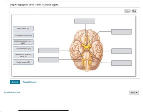
Table of Contents
Drag the Appropriate Labels to Their Respective Targets: A Comprehensive Guide to the Facial Nerve
The facial nerve (cranial nerve VII) is a complex structure with a multifaceted role in facial expression, taste sensation, and even tear and saliva production. Understanding its anatomy and function is crucial for medical professionals, students, and anyone interested in the intricacies of the human nervous system. This comprehensive guide will delve into the detailed anatomy of the facial nerve, its branching pathways, and the clinical implications of its dysfunction. We will explore the different targets associated with the facial nerve and guide you through accurately labeling its components. Think of this as your interactive anatomy lesson – let's drag those labels into place!
The Intricate Anatomy of the Facial Nerve: A Detailed Look
The facial nerve's journey begins deep within the brainstem, emerging from the pons. Its path is anything but straightforward, characterized by several key landmarks and branches that contribute to its complex functionality. Mastering these anatomical details is essential to accurately label its various components.
1. Intracranial Course: From Brainstem to Internal Acoustic Meatus
The facial nerve's journey begins at the facial colliculus, a prominence located on the floor of the fourth ventricle. From here, it enters the internal acoustic meatus, a bony canal shared with the vestibulocochlear nerve (cranial nerve VIII). This shared pathway is significant clinically, as lesions in this region can affect both hearing and facial nerve function.
2. Facial Canal and the Labyrinthine Segment: Navigating the Temporal Bone
Within the temporal bone, the facial nerve follows a winding course through the facial canal. This portion is further divided into segments, with the labyrinthine segment being the initial part. It's important to note the close proximity of the facial nerve to the middle ear structures during this stage. Infections or trauma in this area can easily lead to facial nerve paralysis.
3. Tympanic Segment and the Mastoid Segment: A Journey Through the Middle Ear and Mastoid Process
The tympanic segment passes through the middle ear cavity, running horizontally near the oval window and the stapes. Its close relationship with these structures makes it vulnerable to injury during middle ear surgeries. Subsequently, the nerve enters the mastoid segment, traversing the mastoid portion of the temporal bone. This segment is often the site of surgical approaches to the facial nerve, particularly in cases of decompression or repair.
4. Stylomastoid Foramen: Emerging from the Temporal Bone
After navigating the mastoid process, the facial nerve finally exits the temporal bone through the stylomastoid foramen. This represents a significant transition point, marking the nerve's entry into the extracranial space.
5. Extracranial Course: Branching and Innervation
Once outside the temporal bone, the facial nerve enters the parotid gland, dividing into its five major terminal branches. This is the most clinically relevant portion for understanding facial expression. Accurate labeling of these branches is essential for understanding the specific muscles they innervate:
- Temporal branch: Innervates the frontal and orbicularis oculi muscles, responsible for eyebrow elevation and eye closure.
- Zygomatic branch: Innervates the muscles of the cheek, responsible for smiling and cheek elevation.
- Buccal branch: Innervates the muscles of the mouth, responsible for lip movements and smiling.
- Marginal mandibular branch: Innervates the muscles of the lower lip and chin, crucial for lip pursing and frowning.
- Cervical branch: Innervates the platysma muscle in the neck, responsible for neck muscle movement.
Clinical Significance: Understanding Facial Nerve Dysfunction
Dysfunction of the facial nerve can manifest in a myriad of ways, depending on the location and extent of the lesion. Accurate diagnosis relies on a thorough understanding of its anatomy and the resulting clinical picture.
1. Bell's Palsy: A Common Cause of Facial Nerve Paralysis
Bell's palsy is a common cause of unilateral facial nerve paralysis, often characterized by sudden onset of weakness or paralysis of facial muscles. The exact cause remains elusive, but it's thought to be associated with viral infections or inflammation of the facial nerve within the facial canal.
2. Ramsay Hunt Syndrome: Herpes Zoster Oticus
Ramsay Hunt syndrome is a more complex condition caused by the reactivation of the varicella-zoster virus within the geniculate ganglion, a part of the facial nerve. It's characterized by facial paralysis, ear pain, and vesicles in the ear canal.
3. Acoustic Neuroma: A Tumor Affecting the Facial and Vestibulocochlear Nerves
Acoustic neuroma, also known as vestibular schwannoma, is a benign tumor that arises from the Schwann cells of the vestibulocochlear nerve. However, its location in the internal acoustic meatus often leads to compression of the adjacent facial nerve, resulting in facial weakness or paralysis.
Interactive Exercises: Testing Your Knowledge
Now that we've reviewed the anatomy and clinical correlations, let's put your knowledge to the test with some interactive exercises. Imagine you are presented with a diagram of the facial nerve, and you need to drag and drop labels onto their corresponding anatomical structures.
Exercise 1: Identifying the Intracranial and Intraosseous Segments
You are given a diagram showing the facial nerve from its origin in the brainstem to its exit from the stylomastoid foramen. Drag and drop the following labels onto their correct locations:
- Facial Colliculus
- Internal Acoustic Meatus
- Labyrinthine Segment
- Tympanic Segment
- Mastoid Segment
- Stylomastoid Foramen
Exercise 2: Labeling the Extracranial Branches
A diagram of the extracranial facial nerve is presented, showing its five major branches. Drag and drop the following labels onto their correct locations:
- Temporal Branch
- Zygomatic Branch
- Buccal Branch
- Marginal Mandibular Branch
- Cervical Branch
Exercise 3: Clinical Scenarios
For each clinical scenario below, identify the most likely location of the facial nerve lesion:
- Scenario A: Patient presents with unilateral facial paralysis, with weakness affecting all branches of the facial nerve. There is no associated hearing loss.
- Scenario B: Patient presents with facial paralysis and hearing loss on the same side.
- Scenario C: Patient presents with facial paralysis, severe ear pain, and vesicles in the ear canal.
By completing these exercises, you will reinforce your understanding of the facial nerve's intricate anatomy and its clinical relevance.
Conclusion: Mastering the Facial Nerve
The facial nerve, with its complex anatomy and crucial functions, represents a fascinating area of study within the human nervous system. A thorough understanding of its various components, branching patterns, and clinical implications is crucial for healthcare professionals. The exercises provided here serve as a tool for solidifying your knowledge and building a robust foundation in facial nerve anatomy and neurology. Remember to practice consistently, and you'll soon be a master of dragging those labels to their respective targets!
Latest Posts
Latest Posts
-
Differential White Blood Cell Count Data Table Answers
Mar 19, 2025
-
Practice Phylogenetic Trees 1 Answer Key
Mar 19, 2025
-
The Atoms Family Atomic Math Challenge
Mar 19, 2025
-
Boy I Cant Believe It Is Almost October
Mar 19, 2025
-
Niosh Alerts Disclose New Observations In Regard To
Mar 19, 2025
Related Post
Thank you for visiting our website which covers about Drag The Appropriate Labels To Their Respective Targets. Facial Nerve . We hope the information provided has been useful to you. Feel free to contact us if you have any questions or need further assistance. See you next time and don't miss to bookmark.
