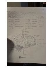
Figure 7-3 Is A Diagram Of The Right Lateral
Onlines
Apr 03, 2025 · 6 min read

Table of Contents
Decoding Figure 7-3: A Deep Dive into the Right Lateral View of the Human Anatomy
Figure 7-3, a diagram depicting the right lateral view of the human anatomy, serves as a fundamental visual tool for understanding the complex arrangement of organs, bones, and muscles within the human body. This article will comprehensively analyze the information likely presented in such a diagram, discussing the key anatomical structures visible from this perspective and their significance in overall bodily function. We will explore the positional relationships between these structures and delve into the clinical implications of understanding this particular view. Because the specific content of Figure 7-3 is unknown, this article will provide a generalized yet detailed analysis of what one would expect to find in a typical right lateral anatomical diagram.
The Importance of Anatomical Views
Before delving into the specifics, it's crucial to understand the importance of different anatomical views in studying the human body. The human body is three-dimensional, and a single view cannot fully capture its complexity. Different views – anterior (front), posterior (back), lateral (side), superior (top), and inferior (bottom) – provide essential perspectives for understanding the spatial relationships between different structures. The right lateral view, specifically, offers a unique perspective on the body’s organization, highlighting the relative positions of structures that might be obscured in other views. This perspective is invaluable for:
- Understanding spatial relationships: The right lateral view clearly shows the layering of structures, revealing which organs are superficial (closer to the surface) and which are deep (further from the surface).
- Diagnosing and treating injuries: In medical imaging and surgery, understanding the right lateral view is crucial for accurately locating injuries and planning surgical procedures.
- Understanding physiological processes: The spatial arrangement of organs, as seen in this view, directly relates to their functions and interactions within the body.
Key Anatomical Structures Visible in the Right Lateral View (Figure 7-3 Hypothetical Content)
A typical right lateral anatomical diagram (like the hypothetical Figure 7-3) would likely showcase a range of structures, including:
Skeletal System:
- Skull: The lateral view of the skull reveals the temporal bone, parietal bone, zygomatic arch (cheekbone), and mandible (jawbone). These bones protect the brain and provide attachment points for facial muscles.
- Vertebral Column: The spinal column is prominently displayed, showcasing the cervical (neck), thoracic (chest), lumbar (lower back), and sacral (pelvic) vertebrae. The curvature of the spine is clearly visible in this view.
- Rib Cage: The ribs, along with the sternum (breastbone), form the protective rib cage, encompassing the heart and lungs. The right lateral view shows the individual ribs and their articulation with the thoracic vertebrae.
- Pelvis: The pelvic bones – ilium, ischium, and pubis – are clearly seen, forming the foundation of the lower body and providing support for the abdominal organs.
- Upper and Lower Limbs: The humerus (upper arm bone), radius and ulna (forearm bones), femur (thigh bone), tibia and fibula (lower leg bones), and associated joints would be visible. The position and articulation of these bones indicate posture and movement capabilities.
Muscular System:
- Superficial Muscles: Numerous superficial muscles would be represented, showing their origin and insertion points on the bones. These include muscles of the back, such as the trapezius and latissimus dorsi; muscles of the shoulder and arm, such as the deltoids, biceps brachii, and triceps brachii; and muscles of the leg, such as the gluteus maximus, quadriceps, hamstrings, and gastrocnemius.
- Deep Muscles: Depending on the detail of Figure 7-3, some deeper muscle layers might also be illustrated, particularly those relevant to posture and movement.
Organ Systems:
- Respiratory System: The lungs, particularly the right lung, would be partially visible, showing their location within the rib cage. The diaphragm, the muscle separating the thoracic and abdominal cavities, would also be represented.
- Cardiovascular System: A portion of the heart might be visible, though it largely resides more medially (towards the midline) and would be better appreciated in an anterior view. Major blood vessels like the aorta and vena cava might be partially visible depending on the level of detail.
- Digestive System: Parts of the liver, stomach, intestines, and possibly the spleen might be partially visible in the right lateral view, depending on the level of detail in Figure 7-3. These organs' relative positions within the abdominal cavity would be highlighted.
- Urinary System: Depending on the level of detail, the right kidney and perhaps part of the ureter might be illustrated.
Clinical Significance of Understanding the Right Lateral View
A strong understanding of the right lateral view, as depicted in a diagram like Figure 7-3, holds significant clinical relevance:
- Trauma Assessment: In cases of trauma, the right lateral view is crucial for assessing injuries to the ribs, spine, and internal organs. This view can help identify fractures, internal bleeding, and other life-threatening conditions.
- Surgical Planning: Surgeons rely heavily on anatomical diagrams to plan surgical approaches. The right lateral view assists in understanding the spatial relationships between structures and aids in determining the safest and most effective surgical techniques.
- Radiological Interpretation: Medical imaging techniques, such as X-rays and CT scans, often provide images from multiple angles, including the right lateral view. Understanding this view enables healthcare professionals to accurately interpret medical images and diagnose pathologies.
- Physical Therapy and Rehabilitation: The right lateral view is vital for physical therapists to understand musculoskeletal alignment, identify muscle imbalances, and create effective treatment plans.
Beyond the Diagram: Connecting Figure 7-3 to Real-World Applications
The information provided by Figure 7-3, whether a simple illustration or a highly detailed anatomical chart, isn't just theoretical knowledge. It has tangible implications across various healthcare professions:
- Emergency Medicine: Paramedics and emergency room physicians must quickly assess patients' injuries using their knowledge of anatomy, including the right lateral view. This enables them to prioritize treatment and stabilize patients effectively.
- Sports Medicine: Athletes are susceptible to various injuries. Understanding the right lateral view is essential for diagnosing and treating these injuries, ensuring athletes recover quickly and safely.
- Geriatric Care: As individuals age, their anatomical structures and functions change. Understanding the right lateral view can aid in diagnosing age-related conditions and planning appropriate care.
- Pediatric Care: Children's anatomy differs from adults'. Knowing the right lateral view in children's anatomy aids pediatricians in diagnosing and treating pediatric conditions.
Enhancing Understanding: Active Learning Strategies
To maximize learning from Figure 7-3 or any similar anatomical diagram, consider these active learning strategies:
- Labeling Exercises: Practice labeling the structures shown in the diagram. This reinforces your understanding of their names and locations.
- Cross-Referencing: Compare the right lateral view with other anatomical views (anterior, posterior, etc.) to gain a more holistic understanding of the body's three-dimensional structure.
- Clinical Case Studies: Apply your knowledge of the right lateral view to clinical case studies. This helps you connect theoretical knowledge with real-world applications.
- 3D Models and Interactive Resources: Utilize 3D anatomical models or interactive software to explore the body from multiple perspectives and enhance spatial understanding.
Conclusion: The Power of Visualization in Anatomy
Figure 7-3, and similar diagrams showcasing the right lateral view of the human anatomy, represent an indispensable tool for understanding the human body. By meticulously studying this perspective, healthcare professionals and students alike can gain a deeper understanding of the intricate relationships between various anatomical structures, leading to improved diagnosis, treatment, and overall patient care. Remember to utilize active learning strategies to maximize your comprehension and apply this knowledge effectively. The ability to visualize and interpret anatomical views, like the right lateral perspective, is fundamental to success in any healthcare-related field.
Latest Posts
Latest Posts
-
Pudieron Terminar El Trabajo Haber Empezado Having Begun A Tiempo
Apr 04, 2025
-
Pre Lab Video Coaching Activity Muscle Contraction
Apr 04, 2025
-
In 2014 85 Percent Of Households
Apr 04, 2025
-
Chapter 1 Test Form A What Is Economics
Apr 04, 2025
-
Pride And Prejudice Summary Chapter 13
Apr 04, 2025
Related Post
Thank you for visiting our website which covers about Figure 7-3 Is A Diagram Of The Right Lateral . We hope the information provided has been useful to you. Feel free to contact us if you have any questions or need further assistance. See you next time and don't miss to bookmark.
