Identify The Phase Of Mitosis Shown In The Highlighted Cell
Onlines
Mar 20, 2025 · 7 min read
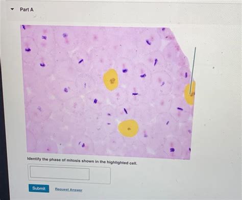
Table of Contents
- Identify The Phase Of Mitosis Shown In The Highlighted Cell
- Table of Contents
- Identify the Phase of Mitosis Shown in the Highlighted Cell: A Comprehensive Guide
- The Five Stages of Mitosis: A Detailed Overview
- 1. Prophase: The Initial Stage of Chromosome Condensation
- 2. Prometaphase: Microtubule Attachment and Chromosome Movement
- 3. Metaphase: Chromosomes Align at the Metaphase Plate
- 4. Anaphase: Sister Chromatids Separate
- 5. Telophase: Chromosomes Decondense and Nuclei Reform
- Identifying the Phase: A Practical Approach
- Case Studies: Analyzing Microscopic Images
- Advanced Considerations and Potential Challenges
- Conclusion: Mastering Mitosis Identification
- Latest Posts
- Latest Posts
- Related Post
Identify the Phase of Mitosis Shown in the Highlighted Cell: A Comprehensive Guide
Understanding the phases of mitosis is crucial for comprehending cell division and its role in growth, repair, and asexual reproduction. This article provides a detailed explanation of the mitotic phases, focusing on how to accurately identify the phase depicted in a given microscopic image, particularly when a specific cell is highlighted. We will delve into the characteristic features of each stage – prophase, prometaphase, metaphase, anaphase, and telophase – equipping you with the knowledge to confidently identify the phase of mitosis shown in any highlighted cell.
The Five Stages of Mitosis: A Detailed Overview
Mitosis is a fundamental process in eukaryotic cells, ensuring the accurate duplication and segregation of chromosomes into two identical daughter cells. This complex process is divided into five distinct stages, each characterized by specific chromosomal and cellular events:
1. Prophase: The Initial Stage of Chromosome Condensation
Prophase marks the beginning of mitosis. During this phase, several key events occur:
-
Chromosome Condensation: The chromatin fibers, which are long, thin strands of DNA and protein, begin to condense and coil tightly, forming visible chromosomes. Each chromosome now consists of two identical sister chromatids joined at the centromere. This condensation is a crucial step, making the chromosomes easier to manage and segregate during subsequent phases. You'll see distinct, rod-like structures under the microscope.
-
Nuclear Envelope Breakdown: The nuclear envelope, the membrane surrounding the nucleus, begins to fragment and disappear. This allows the chromosomes access to the mitotic spindle, a structure responsible for chromosome movement. The disappearance of the nuclear envelope is a definitive visual cue in microscopic images.
-
Spindle Fiber Formation: The centrosomes, which contain centrioles (in animal cells), migrate to opposite poles of the cell. Between the centrosomes, the mitotic spindle begins to form. This spindle is composed of microtubules, protein fibers that will attach to the chromosomes and guide their movement. Observing the centrosomes and the emerging spindle fibers is key to identifying prophase.
2. Prometaphase: Microtubule Attachment and Chromosome Movement
Prometaphase is a transitional phase between prophase and metaphase. It's characterized by the following:
-
Microtubule Attachment to Kinetochores: Kinetochores, protein structures located at the centromere of each chromosome, become attached to the microtubules of the mitotic spindle. These microtubules, called kinetochore microtubules, are crucial for moving the chromosomes to the metaphase plate. Identifying the attachment of microtubules to kinetochores is a hallmark of prometaphase.
-
Chromosome Movement: The chromosomes begin to move toward the center of the cell, although they are not yet aligned precisely. This movement is driven by the dynamic interactions between the kinetochore microtubules and the motor proteins associated with the kinetochores. Observing the movement and dynamic nature of chromosomes is essential for distinguishing prometaphase from other stages.
-
Continued Spindle Fiber Formation: The spindle fibers continue to elongate and expand, filling the space within the cell. This further defines the cell's preparation for the metaphase alignment.
3. Metaphase: Chromosomes Align at the Metaphase Plate
Metaphase is characterized by the precise alignment of chromosomes at the metaphase plate, an imaginary plane equidistant from the two poles of the cell. This alignment is a critical checkpoint, ensuring that each daughter cell receives a complete set of chromosomes.
-
Chromosome Alignment: All chromosomes are arranged along the metaphase plate, with their centromeres aligned precisely in the center. This meticulous arrangement is a defining feature of metaphase, and easily recognizable in microscopic images. The chromosomes appear condensed and neatly lined up.
-
Spindle Fiber Attachment: Each chromosome is attached to microtubules from both poles of the cell, ensuring that sister chromatids will be pulled apart equally during anaphase. The symmetrical arrangement of spindle fibers is a key indicator of metaphase.
-
Spindle Checkpoint Activation: A critical checkpoint mechanism ensures that all chromosomes are properly attached to the spindle fibers before proceeding to anaphase. This checkpoint prevents errors in chromosome segregation that can lead to aneuploidy (abnormal chromosome number).
4. Anaphase: Sister Chromatids Separate
Anaphase is the shortest phase of mitosis, characterized by the separation of sister chromatids and their movement toward opposite poles of the cell.
-
Sister Chromatid Separation: The centromeres of each chromosome split, separating the sister chromatids. These newly separated chromatids are now considered individual chromosomes. This splitting is a dramatic visual change and a defining characteristic of anaphase.
-
Chromosome Movement: The separated chromosomes are pulled toward opposite poles of the cell by the shortening of the kinetochore microtubules. This movement is facilitated by motor proteins and results in a distinct V-shape of the chromosomes as they move.
-
Spindle Elongation: The cell elongates as the poles move further apart. This elongation is driven by the lengthening of the non-kinetochore microtubules (polar microtubules), further contributing to the distinct visual cues of anaphase.
5. Telophase: Chromosomes Decondense and Nuclei Reform
Telophase is the final phase of mitosis, marked by the reversal of many prophase events. It's characterized by:
-
Chromosome Decondensation: The chromosomes begin to uncoil and decondense, returning to their chromatin form. This process is the inverse of what happened in prophase. The chromosomes become less visible under the microscope.
-
Nuclear Envelope Reformation: The nuclear envelope reforms around each set of chromosomes, creating two distinct nuclei. The appearance of two separate nuclei is a strong indicator of telophase.
-
Spindle Fiber Disassembly: The mitotic spindle disassembles, its microtubules breaking down. The cellular structures are reorganized in preparation for cytokinesis.
-
Cytokinesis: While technically not part of mitosis, cytokinesis usually overlaps with telophase. This process divides the cytoplasm, creating two separate daughter cells, each with a complete set of chromosomes and organelles. The formation of a cleavage furrow (in animal cells) or a cell plate (in plant cells) signifies cytokinesis.
Identifying the Phase: A Practical Approach
To accurately identify the phase of mitosis shown in a highlighted cell, systematically examine the following features:
-
Chromosome Condensation: Are the chromosomes highly condensed (prophase, metaphase, anaphase) or decondensed (telophase)? Intermediate condensation suggests prometaphase.
-
Nuclear Envelope: Is the nuclear envelope intact (prophase, early prometaphase), partially broken down (late prometaphase), or completely absent (metaphase, anaphase)?
-
Chromosome Alignment: Are the chromosomes randomly distributed (prophase, prometaphase), aligned at the metaphase plate (metaphase), or moving toward the poles (anaphase)?
-
Sister Chromatids: Are sister chromatids still joined (prophase, prometaphase, metaphase) or separated (anaphase)?
-
Spindle Fibers: Are spindle fibers visible, and if so, are they attached to chromosomes (prometaphase, metaphase, anaphase)?
Case Studies: Analyzing Microscopic Images
Let's consider hypothetical scenarios to illustrate the identification process. Remember, these are simplified examples. Real microscopic images may contain additional complexities.
Scenario 1: The highlighted cell shows highly condensed chromosomes aligned at the center of the cell. The nuclear envelope is absent, and spindle fibers are clearly visible, attached to the chromosomes. The sister chromatids are still joined. This indicates Metaphase.
Scenario 2: The highlighted cell exhibits condensed chromosomes moving toward opposite poles. The sister chromatids are separated. The nuclear envelope is absent, and the spindle fibers are visible. This indicates Anaphase.
Scenario 3: The highlighted cell shows partially condensed chromosomes scattered within a cell with a partially disassembled nuclear envelope. Microtubules are extending towards the chromosomes. This indicates Prometaphase.
Scenario 4: The highlighted cell displays elongated chromosomes that are beginning to decondense. Two distinct sets of chromosomes are forming, and the nuclear envelopes are reforming. This suggests Telophase.
Scenario 5: The highlighted cell shows condensed chromosomes that are not yet aligned, and the nuclear envelope is breaking down. Spindle fibres are starting to form. This is indicative of Prophase.
Advanced Considerations and Potential Challenges
While the above descriptions provide a solid framework, real-world applications can present challenges. Microscopic images might be of lower resolution, the staining may not be ideal, or the cells might be at a transitional stage between phases. In these cases, careful observation, combined with understanding the dynamic nature of mitosis, is critical.
Furthermore, slight variations in appearance can occur due to differences in cell type and experimental conditions. Always refer to the specific context of the image and any accompanying information.
Conclusion: Mastering Mitosis Identification
Identifying the phase of mitosis in a highlighted cell requires a thorough understanding of the characteristic features of each phase. By carefully examining chromosome condensation, nuclear envelope integrity, chromosome alignment, sister chromatid separation, and spindle fiber arrangement, you can accurately determine the stage of mitosis depicted in any given microscopic image. Remember to consider the context and potential limitations of the image itself. With practice and a systematic approach, you'll develop the skill to confidently identify the phase of mitosis, enhancing your comprehension of this fundamental biological process.
Latest Posts
Latest Posts
-
Nr 507 Week 1 Case Study
Mar 21, 2025
-
Activities Such As Purchasing Raw Materials Are Considered Activities
Mar 21, 2025
-
Rn 3 0 Clinical Judgement Practice 2
Mar 21, 2025
-
Which Of The Following Best Describes Bluesnarfing
Mar 21, 2025
-
Studies Published And Unpublished Are Included
Mar 21, 2025
Related Post
Thank you for visiting our website which covers about Identify The Phase Of Mitosis Shown In The Highlighted Cell . We hope the information provided has been useful to you. Feel free to contact us if you have any questions or need further assistance. See you next time and don't miss to bookmark.
