Label The Photomicrograph Of Thin Skin
Onlines
Mar 13, 2025 · 6 min read
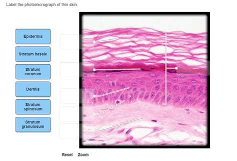
Table of Contents
Labeling a Photomicrograph of Thin Skin: A Comprehensive Guide
Identifying the various layers and structures within a photomicrograph of thin skin (also known as glabrous skin) requires a keen understanding of histology. This detailed guide provides a comprehensive walkthrough, covering essential structures and their characteristics, enabling you to accurately label your own photomicrographs. We will move from the superficial to the deeper layers, highlighting key identifying features and potential points of confusion.
Understanding Thin Skin vs. Thick Skin
Before diving into labeling, it's crucial to understand the differences between thin and thick skin. Thin skin covers most of the body, characterized by the absence of a stratum lucidum, a layer present in thick skin found on the palms and soles. This distinction significantly impacts the appearance of the photomicrograph. While both types share fundamental layers, the relative thicknesses and cellular arrangements differ. This guide focuses specifically on the characteristics of thin skin.
The Epidermis: Layers of Protection
The epidermis, the outermost layer, is avascular and consists of stratified squamous epithelium. It's crucial to correctly identify the following layers:
1. Stratum Corneum: The Protective Barrier
This is the outermost layer, comprising numerous layers of dead, keratinized cells called corneocytes. These cells are flattened and lack nuclei, appearing as anucleated, eosinophilic (pink-staining) scales in the photomicrograph. Look for a densely packed, somewhat homogenous appearance. This layer provides a crucial waterproof barrier and protection against environmental insults.
2. Stratum Granulosum: Granular Layer
Beneath the stratum corneum, the stratum granulosum is characterized by the presence of keratohyalin granules. These granules appear as dark-staining basophilic (blue-staining) structures within the cells. The cells themselves are typically flattened and show signs of beginning keratinization. The presence of keratohyalin granules is a key identifying feature of this layer.
3. Stratum Spinosum: The Prickly Layer
The stratum spinosum is a thicker layer with cells displaying prominent intercellular bridges, giving it a "prickly" appearance. These bridges are actually desmosomes, cell junctions that hold the cells tightly together. The cells themselves are polyhedral (many-sided) and may show some signs of keratinization. Look for the characteristic spiny appearance caused by the numerous desmosomes.
4. Stratum Basale: The Germinative Layer
The deepest layer of the epidermis, the stratum basale, is a single layer of columnar or cuboidal cells. These cells are actively dividing and responsible for the continuous renewal of the epidermis. They are often located adjacent to the basement membrane, separating the epidermis from the dermis. You may observe mitotic figures (cells undergoing division) within this layer.
The Dermis: Connective Tissue Support
The dermis, beneath the epidermis, is a thick layer of connective tissue providing structural support and containing various structures. Key components to identify include:
1. Papillary Layer: Dermal Papillae
This is the superficial layer of the dermis, characterized by finger-like projections called dermal papillae. These papillae interdigitate with the epidermis, increasing the surface area for nutrient exchange and strengthening the adhesion between the two layers. Look for these upward projections extending towards the epidermis. They often contain capillaries and Meissner's corpuscles (discussed later).
2. Reticular Layer: Dense Irregular Connective Tissue
This is the deeper, thicker layer of the dermis, composed of dense irregular connective tissue. It contains collagen and elastic fibers arranged in a complex network, providing strength and elasticity to the skin. This layer appears denser and less organized than the papillary layer. You might observe thicker collagen bundles and fewer cells compared to the papillary layer.
Appendages of the Skin: Structures within the Dermis
Several important structures are embedded within the dermis:
1. Hair Follicles: Hair Growth Units
Hair follicles are tubular invaginations of the epidermis extending deep into the dermis, sometimes reaching the hypodermis. They are responsible for hair growth and consist of several layers, including the inner root sheath and the outer root sheath. Identify the follicle's shape and location within the dermis. The hair shaft itself may be present in the section.
2. Sebaceous Glands: Oil Production
These glands are typically associated with hair follicles and secrete sebum, an oily substance that lubricates the skin and hair. Sebaceous glands appear as clusters of cells with a foamy or vacuolated cytoplasm. They may be located near the hair follicle.
3. Sweat Glands (Eccrine): Thermoregulation
Eccrine sweat glands are coiled tubular glands responsible for thermoregulation. They are typically located deeper within the dermis or even extending into the hypodermis. They are more difficult to identify in thin skin sections than in thick skin sections, often appearing as small, coiled structures.
4. Sensory Receptors: Meissner's Corpuscles and Pacinian Corpuscles
Meissner's corpuscles, responsible for light touch sensation, are located in the dermal papillae. They appear as elongated, oval structures. Pacinian corpuscles, sensitive to deep pressure and vibration, are larger and found deeper within the dermis and hypodermis. They often appear as concentric lamellae, although these might be less apparent in thin sections. Their presence varies significantly depending on the location of the skin sample.
5. Blood Vessels: Nutrient Supply
Blood vessels are essential for supplying nutrients and oxygen to the skin. Capillaries are particularly abundant in the papillary layer, supplying the epidermis and other structures within the dermis. Arteries and veins of larger caliber may also be visible in the deeper dermis.
Labeling Your Photomicrograph: A Step-by-Step Guide
- Identify the Epidermis: Begin by identifying the epidermis, the outermost layer. It is characterized by stratified squamous epithelium.
- Layer by Layer: Carefully delineate the strata of the epidermis (corneum, granulosum, spinosum, basale) based on the cellular characteristics discussed above.
- Locate the Basement Membrane: The basement membrane separates the epidermis from the dermis.
- Distinguish the Dermis: Identify the dermis, composed of connective tissue. Note the papillary and reticular layers.
- Find Appendages: Search for hair follicles, sebaceous glands, and sweat glands within the dermis.
- Identify Sensory Receptors: Look for Meissner's corpuscles in the dermal papillae and potentially Pacinian corpuscles deeper in the dermis.
- Label Blood Vessels: Identify and label the blood vessels present in the dermis.
- Use Labels Clearly: Use clear, concise labels with arrows pointing to the specific structures.
Potential Challenges and Tips
- Stain Variation: The staining intensity might differ depending on the histological staining technique employed (e.g., H&E). This can affect the visibility of certain structures.
- Section Orientation: The orientation of the tissue section can impact the visibility of certain structures. A cross-section might reveal different features than a longitudinal section.
- Artifact Recognition: Be aware of potential artifacts during tissue processing that may be misinterpreted as structures.
By following this guide and carefully analyzing the characteristics of each layer and structure, you can accurately label a photomicrograph of thin skin. Remember to consistently refer to histological textbooks and online resources for additional visual aids and detailed explanations. Practice makes perfect; the more photomicrographs you label, the better you will become at identifying these structures. Good luck!
Latest Posts
Latest Posts
-
Lesson 2 Eggzactly M1 67 Answer Key
Mar 14, 2025
-
Separation Of The Components Of A Mixture Pre Lab Answers
Mar 14, 2025
-
The Inzinzebu Bandits Are Harassing The Good Merchants In
Mar 14, 2025
-
Introduction To Acids And Bases Webquest Answer Key
Mar 14, 2025
-
A Newly Formed From Must Decide
Mar 14, 2025
Related Post
Thank you for visiting our website which covers about Label The Photomicrograph Of Thin Skin . We hope the information provided has been useful to you. Feel free to contact us if you have any questions or need further assistance. See you next time and don't miss to bookmark.
