Label The Tissue Types Illustrated Here
Onlines
Apr 06, 2025 · 6 min read
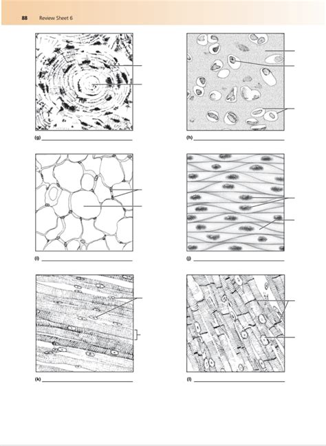
Table of Contents
Label the Tissue Types Illustrated Here: A Comprehensive Guide to Histology
Histology, the microscopic study of tissues, is fundamental to understanding the structure and function of the human body. This article serves as a comprehensive guide to identifying and labeling various tissue types, providing detailed descriptions and illustrative examples. We’ll cover the four primary tissue types: epithelial, connective, muscle, and nervous tissue, exploring their subtypes and key characteristics. Mastering tissue identification is crucial for students in biology, medicine, and related fields.
I. Epithelial Tissue: Covering and Lining
Epithelial tissue forms the linings of organs and body cavities, and also constitutes glands. Its defining characteristics are cellularity (tightly packed cells), specialized contacts (cell junctions), polarity (apical and basal surfaces), support by connective tissue (basement membrane), avascularity (lack of blood vessels), and regeneration.
A. Classifying Epithelial Tissues
Epithelial tissues are classified based on two criteria:
- Cell shape: Squamous (flattened), cuboidal (cube-shaped), columnar (tall and column-shaped).
- Cell arrangement: Simple (single layer), stratified (multiple layers), pseudostratified (appears stratified but is a single layer).
B. Examples of Epithelial Tissues:
1. Simple Squamous Epithelium: Characterized by a single layer of flattened cells. It's found where diffusion or filtration occurs, such as in the lining of blood vessels (endothelium) and alveoli of the lungs. Function: Facilitates rapid diffusion and filtration.
2. Simple Cuboidal Epithelium: Composed of a single layer of cube-shaped cells. It's found in glands and ducts, as well as in the lining of kidney tubules. Function: Secretion and absorption.
3. Simple Columnar Epithelium: Features a single layer of tall, column-shaped cells. Often contains goblet cells (producing mucus). It lines the digestive tract. Function: Secretion and absorption; goblet cells provide lubrication. Some types possess cilia for movement.
4. Pseudostratified Columnar Epithelium: Appears layered but is actually a single layer of cells with varying heights. Often ciliated (e.g., in the trachea). Function: Secretion (e.g., mucus) and movement of mucus via cilia.
5. Stratified Squamous Epithelium: Multiple layers of cells, with flattened cells at the surface. Found in areas subject to abrasion, such as the epidermis of the skin and lining of the esophagus. Function: Protection against abrasion. Keratinized stratified squamous epithelium (like skin) is resistant to water loss.
6. Stratified Cuboidal Epithelium: Rare; found in some ducts of large glands. Function: Protection and secretion.
7. Stratified Columnar Epithelium: Also rare; found in some ducts of large glands and parts of the male urethra. Function: Protection and secretion.
8. Transitional Epithelium: Specialized epithelium found in the urinary system. Its cells can change shape, allowing for distension (stretching) of the organ. Function: Allows for stretching of the urinary organs.
II. Connective Tissue: Support and Binding
Connective tissue is the most abundant and widely distributed tissue type. Its primary functions are binding and support, protection, insulation, and transportation (blood). Connective tissues are characterized by having abundant extracellular matrix (ECM), which consists of ground substance and fibers.
A. Components of Connective Tissue:
- Ground substance: A gel-like substance filling the space between cells and fibers.
- Fibers: Collagen fibers (strong and flexible), elastic fibers (stretchy), and reticular fibers (fine, branching fibers).
- Cells: Various types of cells, including fibroblasts (produce ECM), chondrocytes (cartilage cells), osteocytes (bone cells), adipocytes (fat cells), and blood cells.
B. Types of Connective Tissues:
1. Connective Tissue Proper: Loose and dense connective tissues.
- Loose Connective Tissue: Areolar, adipose, and reticular. Areolar tissue is a general packing material, adipose tissue stores fat, and reticular tissue forms the framework of lymphoid organs.
- Dense Connective Tissue: Dense regular (tendons and ligaments), dense irregular (dermis of skin), and elastic (walls of large arteries).
2. Specialized Connective Tissues:
- Cartilage: Hyaline (smooth, found in articular cartilage), elastic (flexible, found in ears), and fibrocartilage (strong, found in intervertebral discs).
- Bone: Compact (dense, forms the outer layer of bones) and spongy (porous, found inside bones).
- Blood: A fluid connective tissue consisting of plasma (ground substance), red blood cells, white blood cells, and platelets.
III. Muscle Tissue: Movement
Muscle tissue is responsible for movement. Three types of muscle tissue exist: skeletal, smooth, and cardiac.
A. Skeletal Muscle Tissue:
- Characteristics: Striated (striped), voluntary (under conscious control), multinucleated (multiple nuclei per cell).
- Function: Movement of bones and other structures.
B. Smooth Muscle Tissue:
- Characteristics: Non-striated (smooth), involuntary (not under conscious control), uninucleated (one nucleus per cell).
- Function: Movement of internal organs (e.g., digestion, blood vessel constriction).
C. Cardiac Muscle Tissue:
- Characteristics: Striated, involuntary, branched cells with intercalated discs (specialized junctions).
- Function: Contraction of the heart.
IV. Nervous Tissue: Communication
Nervous tissue is specialized for communication. It consists of neurons (nerve cells) and neuroglia (supporting cells).
A. Neurons:
- Characteristics: Excitable cells that transmit electrical signals. They have a cell body (soma), dendrites (receive signals), and an axon (transmits signals).
- Function: Rapid communication throughout the body.
B. Neuroglia:
- Characteristics: Supporting cells that provide nutrients, insulation, and protection to neurons.
- Function: Maintain the environment for neuron function.
V. Labeling Tissue Types in Microscopic Images:
To accurately label tissue types in microscopic images, you need to carefully observe the following features:
- Cell shape and arrangement: Are the cells flat, cuboidal, or columnar? Are they arranged in a single layer or multiple layers?
- Presence of specialized structures: Are there cilia, microvilli, goblet cells, or intercalated discs?
- Type of extracellular matrix: Is the matrix abundant or sparse? What type of fibers are present?
- Overall tissue organization: How are the cells and matrix organized?
By systematically analyzing these features, you can accurately identify and label the various tissue types presented in a microscopic image. Practice is key to mastering this skill. Regularly reviewing histological slides and comparing them with detailed descriptions will greatly improve your ability to differentiate between various tissue types. Remember to always consult reputable resources and textbooks for further clarification and deeper understanding.
VI. Advanced Techniques in Histology:
Modern histology utilizes advanced techniques to visualize tissues and their components. These include immunohistochemistry (IHC), which uses antibodies to identify specific proteins within tissues; in situ hybridization (ISH), which detects specific DNA or RNA sequences; and electron microscopy, which provides extremely high-resolution images of tissue ultrastructure. Understanding these advanced techniques helps to further refine the identification and characterization of tissue types, revealing details not apparent with traditional light microscopy.
VII. Clinical Significance:
Accurate identification of tissue types is crucial in various clinical settings. Histopathological analysis of tissue samples is vital for diagnosing diseases like cancer, inflammation, and other conditions. The characteristics of tissues observed under the microscope help pathologists determine the nature of a disease, its stage of progression, and potential treatment strategies. Therefore, a comprehensive understanding of histology is essential for accurate diagnosis and effective medical care.
This comprehensive guide provides a strong foundation for identifying and labeling tissue types. Consistent study and practice with microscopic images will enhance your proficiency in histology and its applications in various scientific and medical fields. Remember that this is a vast subject; continuous learning and exploration are essential for mastery.
Latest Posts
Latest Posts
-
Chapter 2 Skills And Applications Answer Key
Apr 07, 2025
-
Which Of The Following M And A Transaction Equations Is Correct
Apr 07, 2025
-
Decidimos Venir A Este Restaurante Porque Mi Jefe
Apr 07, 2025
-
Martin Puts Two Bowls Of Fruit
Apr 07, 2025
-
For Stemi Patients Which Best Describes
Apr 07, 2025
Related Post
Thank you for visiting our website which covers about Label The Tissue Types Illustrated Here . We hope the information provided has been useful to you. Feel free to contact us if you have any questions or need further assistance. See you next time and don't miss to bookmark.
