Onion Cell Mitosis Answer Key Pdf
Onlines
Mar 06, 2025 · 6 min read
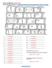
Table of Contents
Decoding the Onion Cell Mitosis: A Comprehensive Guide
Finding a readily available "onion cell mitosis answer key PDF" might prove elusive. The process of mitosis, particularly in onion root tip cells, is best understood through hands-on observation and analysis, not solely through pre-made answer keys. However, this comprehensive guide will equip you with the knowledge and understanding necessary to accurately identify and interpret the stages of mitosis in onion root tip cells, effectively rendering an "answer key" unnecessary. We'll delve deep into the process, focusing on the visual cues and characteristics of each phase. This detailed understanding will empower you to analyze your own microscopic observations and confidently assess the cell cycle stages.
Understanding Mitosis: The Foundation of Cell Division
Mitosis is a fundamental process in all eukaryotic cells, responsible for cell growth and repair. It's a type of cell division that results in two daughter cells, each having the same number and kind of chromosomes as the parent cell. This precise duplication ensures genetic continuity across generations of cells. The process is remarkably complex and involves several distinct phases:
The Stages of Mitosis: A Detailed Breakdown
Mitosis is further divided into several key stages: prophase, prometaphase, metaphase, anaphase, and telophase. Cytokinesis, the division of the cytoplasm, typically follows telophase. Let's explore each phase in detail:
1. Prophase: The Preparatory Stage
Key Characteristics:
- Chromatin Condensation: The diffuse chromatin fibers condense into visible chromosomes. Each chromosome consists of two identical sister chromatids joined at the centromere. This condensation is crucial for efficient segregation during later stages.
- Nuclear Envelope Breakdown: The nuclear membrane surrounding the nucleus disintegrates, allowing the chromosomes to access the cytoplasm.
- Spindle Formation: The mitotic spindle, a structure made of microtubules, begins to form. This structure plays a vital role in separating the chromosomes.
- Centrosome Duplication & Migration: In animal cells, centrosomes (microtubule-organizing centers) duplicate and migrate to opposite poles of the cell. Plant cells lack centrosomes, but the spindle apparatus still forms.
Microscopic Observation: You'll observe thickened, condensed chromosomes within the cell. The nuclear envelope will appear to be disintegrating or fragmented.
2. Prometaphase: Connecting to the Spindle
Key Characteristics:
- Kinetochore Formation: Protein complexes called kinetochores assemble at the centromeres of each chromosome. These kinetochores serve as attachment points for the spindle microtubules.
- Chromosome Capture: Spindle microtubules attach to the kinetochores, connecting the chromosomes to the spindle poles. This connection is crucial for the precise movement of chromosomes in subsequent stages.
Microscopic Observation: Chromosomes appear more organized and aligned. You might see microtubules extending from the poles towards the chromosomes.
3. Metaphase: Alignment at the Equator
Key Characteristics:
- Chromosomal Alignment: The chromosomes align along the metaphase plate, an imaginary plane equidistant from the two spindle poles. This alignment ensures that each daughter cell receives one copy of each chromosome.
- Spindle Checkpoint: The cell meticulously checks to ensure that all chromosomes are correctly attached to the spindle before proceeding to anaphase. This checkpoint prevents errors in chromosome segregation.
Microscopic Observation: Chromosomes will be clearly visible, arranged in a single line along the cell's equator.
4. Anaphase: Sister Chromatid Separation
Key Characteristics:
- Sister Chromatid Separation: The centromeres divide, and the sister chromatids separate, becoming individual chromosomes.
- Chromosome Movement: The separated chromosomes move towards opposite poles of the cell, guided by the shortening of the spindle microtubules.
Microscopic Observation: You'll observe the chromosomes moving towards opposite ends of the cell. The cell elongates slightly.
5. Telophase: Re-formation and Division
Key Characteristics:
- Chromosome Decondensation: The chromosomes begin to decondense, reverting to their less compact chromatin form.
- Nuclear Envelope Reformation: A nuclear envelope reforms around each set of chromosomes, creating two distinct nuclei.
- Spindle Disassembly: The mitotic spindle disassembles.
Microscopic Observation: Two distinct nuclei will be visible, each with a complete set of chromosomes. The chromosomes appear less condensed.
6. Cytokinesis: Cytoplasmic Division
Key Characteristics:
- Cell Division: The cytoplasm divides, resulting in two separate daughter cells. In plant cells, a cell plate forms, eventually developing into a new cell wall. Animal cells undergo a cleavage furrow.
Microscopic Observation: Two completely separate daughter cells will be visible. You might observe a cell plate (plants) or cleavage furrow (animals).
Onion Root Tip Cells: An Ideal Model
Onion root tip cells are frequently used in educational settings to study mitosis due to their rapid cell division rate and ease of preparation for microscopic observation. The meristematic region, located at the root tip, is where the majority of cell division occurs.
Preparing Onion Root Tip Slides: A Step-by-Step Guide
While a detailed step-by-step guide is beyond the scope of this article, the basic procedure involves:
- Collecting the Root Tip: Carefully remove the root tip from a young onion.
- Fixation: Treat the root tip with a fixative to preserve the cell structure.
- Hydrolysis: This step helps to soften the cell walls and separate the chromosomes.
- Staining: Staining the cells (e.g., with acetocarmine) makes the chromosomes easier to visualize under the microscope.
- Mounting: Carefully mount the stained root tip on a microscope slide.
Identifying Mitosis Stages: Practical Tips and Tricks
Accurate identification of the mitotic stages requires careful observation and a good understanding of the characteristics of each stage.
- Chromosome Condensation: The degree of chromosome condensation is a crucial indicator. Prophase shows increasingly condensed chromosomes. Metaphase shows maximum condensation. Telophase shows decondensed chromosomes.
- Nuclear Envelope: The presence or absence of the nuclear envelope is a clear marker. It's intact in interphase and absent in prometaphase and metaphase.
- Spindle Apparatus: Observing the spindle microtubules attaching to the chromosomes helps identify prometaphase and metaphase.
- Chromosome Alignment: The precise alignment of chromosomes at the metaphase plate is characteristic of metaphase.
- Sister Chromatid Separation: The separation of sister chromatids is a definitive sign of anaphase.
- Cytokinesis: The appearance of a cell plate (plants) or cleavage furrow (animals) signals the final stage of cell division.
Beyond the Visual: Understanding the Significance
Understanding mitosis goes beyond simple identification of phases. It's crucial to grasp the implications of accurate chromosome segregation. Errors during mitosis can lead to aneuploidy (abnormal chromosome number), potentially causing developmental problems or diseases like cancer. The intricate choreography of mitosis underscores the remarkable precision of cellular processes.
Conclusion: Mastering Mitosis Through Observation
While a readily available "onion cell mitosis answer key PDF" might be hard to find, the detailed explanations and visual descriptions provided here should serve as a comprehensive guide. By carefully observing prepared slides and understanding the characteristics of each mitotic stage, you can confidently analyze and interpret the cell division process. This hands-on approach fosters deeper understanding and appreciation for the fundamental process of mitosis in eukaryotic cells. Remember, the key to mastering mitosis lies in thorough observation and a strong grasp of the underlying biological principles.
Latest Posts
Latest Posts
-
Question Almond Joy Draw The Skeletal Structure
Mar 06, 2025
-
Aromas Se Quemen De Placido Olor Lyrics
Mar 06, 2025
-
Ana Y Enrique Piden Unos Refrescos Frios
Mar 06, 2025
-
Exercise Methodology Includes Which Of The Following Exercise Cycle Components
Mar 06, 2025
-
How Did You Get To The Top Of Sao Carlos
Mar 06, 2025
Related Post
Thank you for visiting our website which covers about Onion Cell Mitosis Answer Key Pdf . We hope the information provided has been useful to you. Feel free to contact us if you have any questions or need further assistance. See you next time and don't miss to bookmark.
