Osteochondritis Is An Example Of A
Onlines
Mar 16, 2025 · 8 min read
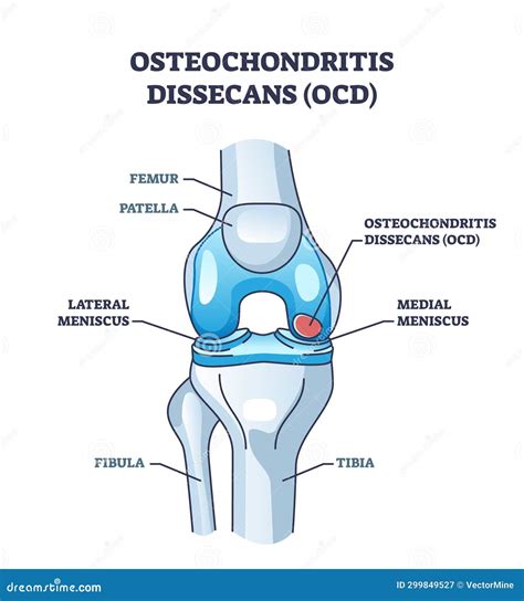
Table of Contents
Osteochondritis Dissecans: An Example of Avascular Necrosis and Joint Injury
Osteochondritis dissecans (OCD) is a fascinating and complex example of avascular necrosis, a condition where bone tissue dies due to a lack of blood supply. It's a relatively common injury, primarily affecting adolescents and young adults, and exemplifies the intricate interplay between bone growth, blood supply, and the biomechanics of joints. Understanding OCD offers valuable insights into the broader field of orthopedic injuries and the delicate balance required for healthy joint function.
What is Osteochondritis Dissecans?
Osteochondritis dissecans is a condition characterized by the separation of a segment of articular cartilage and underlying bone from the rest of the bone. This separation, or detachment, occurs at the joint surface, most commonly in weight-bearing joints like the knee, ankle, elbow, and hip. The affected area, often described as a "lesion," can range in size from a few millimeters to several centimeters. The severity varies considerably, from minor separation to complete detachment of the bone fragment, sometimes leading to it becoming loose within the joint (a “loose body”).
The Underlying Process: Avascular Necrosis
The core pathology behind OCD is avascular necrosis. In simple terms, this means the bone tissue loses its blood supply. This can happen due to a number of reasons, although the exact mechanism isn't fully understood in all cases. The most widely accepted theory suggests that repetitive microtrauma, or small injuries sustained over time, compromises the blood supply to the subchondral bone (the bone directly beneath the cartilage). This compromised blood flow leads to the death of bone cells, resulting in the separation of the cartilage and underlying bone.
Risk Factors Contributing to OCD
Several factors increase the likelihood of developing osteochondritis dissecans:
- Genetics: A predisposition towards OCD can be inherited, suggesting a genetic component to the condition. Family history is a notable risk factor.
- Repetitive Stress: Activities involving repeated high-impact forces or repetitive twisting motions on the joint, such as those seen in athletes participating in sports like gymnastics, basketball, and baseball, substantially increase the risk.
- Growth Spurts: Rapid growth during adolescence can place increased stress on the still-developing bones and joints, making them more vulnerable.
- Trauma: Although not always the sole cause, a single significant injury to the joint, such as a fracture or dislocation, can disrupt the blood supply and initiate the development of OCD.
- Blood Supply Issues: Pre-existing conditions affecting blood flow to the bone, either locally or systemically, can predispose individuals to OCD.
Symptoms of Osteochondritis Dissecans
The symptoms of osteochondritis dissecans can vary depending on the location, size, and severity of the lesion. Some individuals may experience no noticeable symptoms, particularly if the lesion is small and stable. However, more significant lesions can manifest as:
- Pain: Localized pain in the affected joint, often worsened by activity and relieved by rest. The pain can range from mild to severe.
- Swelling: Swelling around the affected joint may be present, especially after activity.
- Stiffness: Stiffness and limited range of motion in the joint are common symptoms.
- Locking or Giving Way: In cases of a loose fragment, the joint might suddenly lock or give way, causing instability and potential falls.
- Clicking or Popping: A clicking or popping sensation within the joint may be felt or heard during movement.
Diagnosis of Osteochondritis Dissecans
Diagnosing OCD typically involves a combination of techniques:
- Physical Examination: A thorough physical examination by an orthopedist is the first step. The doctor will assess the joint's range of motion, palpate for tenderness, and check for any instability.
- Imaging Studies: Imaging plays a crucial role in confirming the diagnosis and determining the severity of the lesion. Commonly used imaging techniques include:
- X-rays: X-rays can reveal the characteristic radiographic features of OCD, such as a flattening or irregularity of the articular surface. They may also show a loose fragment.
- MRI (Magnetic Resonance Imaging): MRI provides the most detailed images of the bone and cartilage, allowing for precise visualization of the lesion and assessment of its stability. It's superior to X-rays in identifying early lesions or subtle changes.
- CT (Computed Tomography) Scan: A CT scan can provide additional information about the bony structure and the integrity of the surrounding tissues. It is less frequently used than MRI in diagnosing OCD.
Treatment Options for Osteochondritis Dissecans
Treatment for osteochondritis dissecans varies depending on factors like the patient's age, the size and location of the lesion, the stability of the fragment, and the presence of symptoms. The goal of treatment is to promote healing and restore joint function.
Non-Surgical Treatment
Non-surgical approaches are often the initial choice, particularly for stable lesions with minimal symptoms:
- Rest and Activity Modification: Limiting activities that stress the affected joint is crucial to allow the lesion to heal. This may involve avoiding certain sports or activities.
- Physical Therapy: Physical therapy plays a vital role in strengthening the muscles surrounding the joint, improving joint stability, and restoring range of motion. Therapeutic exercises and modalities can help manage pain and inflammation.
- Bracing or Immobilization: In some cases, a brace or splint may be used to immobilize the joint and reduce stress on the lesion.
- Pain Management: Over-the-counter pain relievers like ibuprofen or naproxen can help manage pain and inflammation. In more severe cases, stronger prescription pain medication may be necessary.
Surgical Treatment
Surgical intervention may be necessary if the lesion is unstable, symptomatic, or not healing with conservative measures. Several surgical techniques are available:
- Arthroscopy: Arthroscopy is a minimally invasive procedure where small incisions are made to insert a camera and surgical instruments into the joint. This allows for removal of loose fragments, debridement (cleaning up) of damaged cartilage, and promotion of healing.
- Open Surgery: In cases of large lesions or unstable fragments, open surgery may be required. This involves a larger incision to gain access to the joint and perform more extensive procedures, such as bone grafting or fixation of the fragment. This may involve surgical fixation using pins, screws, or other implants to stabilize the bone fragment and promote healing.
Prognosis and Recovery
The prognosis for osteochondritis dissecans is generally good, especially with early diagnosis and appropriate treatment. Most individuals make a full recovery with non-surgical management. However, the recovery process can take several months or even longer, depending on the severity of the lesion and the chosen treatment method. Athletes may need a longer period of rehabilitation to regain their pre-injury level of performance. In some cases, despite treatment, long-term complications such as arthritis may develop later in life, although this is not always the case.
Osteochondritis Dissecans in Different Joints
While OCD can affect various joints, its presentation and management can differ depending on the location:
Osteochondritis Dissecans of the Knee
The knee is the most common site for OCD. The lesions typically occur in the medial femoral condyle (inner part of the thigh bone), though they can also be found in other parts of the knee. Treatment strategies often focus on preserving articular cartilage and restoring joint stability.
Osteochondritis Dissecans of the Ankle
OCD of the ankle is less frequent compared to the knee. The talus (one of the ankle bones) is the most often affected. Treatment may involve either conservative approaches or surgical intervention, depending on the lesion's characteristics and the patient's symptoms.
Osteochondritis Dissecans of the Elbow
The elbow is another common site for OCD, typically affecting the capitellum (the rounded part of the humerus that articulates with the radius). The treatment strategy varies based on the location, size and stability of the lesion.
Osteochondritis Dissecans of the Hip
OCD in the hip is relatively rare. The femoral head is usually the affected area. Conservative treatment is often attempted initially. Surgical intervention may be required for large or unstable lesions.
Living with Osteochondritis Dissecans: Long-Term Outlook
Once the initial treatment phase is complete, it's crucial to follow a structured rehabilitation program. This involves gradually increasing physical activity and strengthening the muscles around the affected joint. Regular physical therapy sessions will help restore range of motion, improve strength and stability, and reduce the risk of re-injury. In some cases, long-term monitoring may be recommended to detect any potential long-term complications. The adherence to the rehabilitation plan is critical for achieving optimal long-term outcomes and minimizing the risk of future issues.
The long-term outlook for individuals with osteochondritis dissecans depends greatly on factors such as the size and location of the lesion, its stability, and the effectiveness of the treatment. Early diagnosis and timely treatment, combined with diligent adherence to rehabilitation protocols, significantly improve the chances of a full recovery and a return to normal activities.
Conclusion: Understanding the Broader Implications
Osteochondritis dissecans, while seemingly a specific condition, provides valuable insights into broader orthopedic principles. It highlights the importance of maintaining adequate blood supply to the bones, the vulnerability of growing bones to repetitive stress and trauma, and the intricate relationship between bone and cartilage health. The multi-faceted approach to diagnosis and treatment underlines the need for a holistic understanding of musculoskeletal injuries, incorporating both conservative and surgical techniques depending on individual circumstances. By studying OCD, we gain a richer comprehension of joint biomechanics, injury mechanisms, and the complex process of healing in the musculoskeletal system. Further research into the exact causes and optimal management strategies for OCD will continue to refine our understanding and improve patient outcomes.
Latest Posts
Latest Posts
-
Elige La Palabra Adecuada Para Completar Las Oraciones Comparativas
Mar 17, 2025
-
You Seem Pleased To Have Successfully Whored Yourself Manacled
Mar 17, 2025
-
Muchas Ninas Jovenes Han Estado A Dieta Terrible
Mar 17, 2025
-
Examples Of Public Data Collected By Law From Physicians Include
Mar 17, 2025
-
Alcohol And Its Effects On The Body Worksheet Answers
Mar 17, 2025
Related Post
Thank you for visiting our website which covers about Osteochondritis Is An Example Of A . We hope the information provided has been useful to you. Feel free to contact us if you have any questions or need further assistance. See you next time and don't miss to bookmark.
