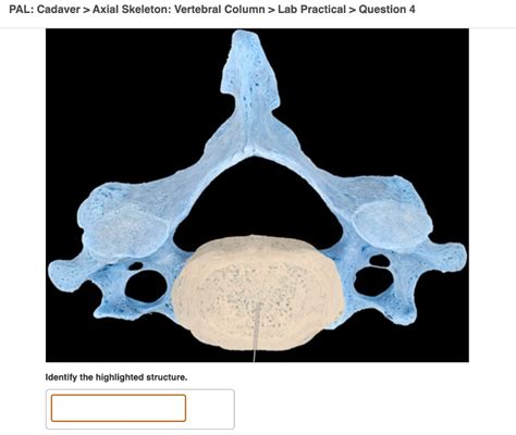
Pal Cadaver Axial Skeleton Vertebral Column Lab Practical Question 4
Onlines
Mar 28, 2025 · 6 min read

Table of Contents
Pal Cadaver Axial Skeleton Vertebral Column Lab Practical Question 4: A Comprehensive Guide
This article delves into the complexities of Question 4 in a typical lab practical focusing on the pal cadaver axial skeleton, specifically the vertebral column. We'll break down the common challenges students face, provide detailed anatomical information, and offer strategies for mastering this crucial area of anatomy. Understanding the vertebral column is fundamental to comprehending human movement, posture, and neurological function.
H2: Understanding the Scope of Question 4
Lab practicals often assess your understanding of anatomical structures through a combination of identification, description, and application. Question 4, centered on the vertebral column within the context of a pal cadaver, likely tests your ability to:
- Identify specific vertebrae: Cervical, thoracic, lumbar, sacrum, and coccyx. This includes recognizing distinguishing features of each vertebral region.
- Describe vertebral characteristics: This includes articulating processes, foramina, and the curvature of the spine.
- Explain functional relationships: How the vertebrae interact with each other, support weight, and protect the spinal cord.
- Analyze pathological changes: (Depending on the complexity of the practical) Identifying potential abnormalities or variations from a "normal" vertebral column.
- Demonstrate proper handling techniques: Showing safe and respectful handling of the pal cadaver.
H2: Detailed Anatomy of the Vertebral Column
The vertebral column, or spine, is a complex structure composed of 33 vertebrae: 7 cervical, 12 thoracic, 5 lumbar, 5 sacral (fused), and 4 coccygeal (fused). Each region exhibits unique characteristics reflecting its functional role.
H3: Cervical Vertebrae (C1-C7)
These are the smallest and most mobile vertebrae, supporting the head and facilitating neck movement. Key features include:
- Atlas (C1): Lacks a body and spinous process; characterized by its ring-like structure with superior and inferior articular facets for articulation with the occipital condyles and axis.
- Axis (C2): Possesses the dens (odontoid process), a unique projection that articulates with the atlas, allowing for rotation of the head.
- Transverse foramina: Present in all cervical vertebrae except C7, these foramina allow passage of the vertebral arteries and veins.
- Bifid spinous processes: Typically, cervical spinous processes are short and bifid (split at the end).
H3: Thoracic Vertebrae (T1-T12)
These vertebrae are larger than cervical vertebrae and articulate with the ribs. Key distinguishing features include:
- Heart-shaped bodies: The vertebral bodies are larger than cervical and increase in size inferiorly.
- Costal facets: These facets on the vertebral bodies and transverse processes articulate with the ribs, forming the costovertebral and costotransverse joints.
- Long, slender spinous processes: These processes point inferiorly.
H3: Lumbar Vertebrae (L1-L5)
These are the largest and strongest vertebrae, bearing most of the body's weight. Characteristics include:
- Large, kidney-shaped bodies: These reflect their weight-bearing function.
- Short, thick, robust spinous processes: These processes project posteriorly.
- Absence of costal facets: These vertebrae do not articulate with ribs.
H3: Sacrum and Coccyx
The sacrum is a triangular bone formed by the fusion of five sacral vertebrae. The coccyx is formed by the fusion of four coccygeal vertebrae, representing the vestigial tailbone. They play a role in pelvic stability and support.
H2: Addressing Common Challenges in Lab Practicals
Students often struggle with differentiating between vertebral regions, identifying subtle anatomical features, and understanding the functional significance of specific structures. Here are some strategies to overcome these challenges:
- Thorough Pre-Lab Preparation: Reviewing anatomical diagrams, atlases, and lecture notes before the practical is crucial. Familiarize yourself with the key features of each vertebral region.
- Utilizing Multiple Learning Resources: Engage with various resources, including textbooks, online anatomy resources, and anatomical models. This multi-modal approach reinforces learning.
- Focusing on Key Distinguishing Features: Create a table summarizing the key differences between cervical, thoracic, and lumbar vertebrae. Focus on size, shape, and presence/absence of specific features (e.g., costal facets, transverse foramina, bifid spinous processes).
- Practical Application: Practice identifying vertebrae on anatomical models and if possible, radiographic images before engaging with the pal cadaver. This will build confidence and familiarity.
- Understanding Functional Anatomy: Connect the anatomical features to their function. For example, understand how the shape of lumbar vertebrae contributes to their weight-bearing role.
H2: Ethical Considerations and Cadaver Handling
Working with a pal cadaver requires utmost respect and adherence to ethical guidelines. Remember:
- Treat the cadaver with dignity: Avoid casual or disrespectful behavior.
- Follow all instructions provided by your instructor: Pay close attention to handling procedures and safety precautions.
- Maintain a professional and respectful environment: Avoid inappropriate conversations or actions.
- Understand the importance of the cadaver's contribution to medical education: Appreciate the generous donation that allows for this valuable learning experience.
H2: Expanding Your Knowledge: Beyond the Basics
To excel in the lab practical, consider broadening your understanding beyond the basics:
- Vertebral Curvatures: Learn about the four normal curvatures of the spine (cervical lordosis, thoracic kyphosis, lumbar lordosis, and sacral kyphosis) and their functional significance. Understand how deviations from these curvatures (e.g., scoliosis) affect posture and function.
- Intervertebral Discs: Study the structure and function of intervertebral discs, including the annulus fibrosus and nucleus pulposus. Understand their role in shock absorption and movement.
- Spinal Ligaments: Familiarize yourself with the major ligaments that support the vertebral column, including the anterior and posterior longitudinal ligaments, ligamentum flavum, and supraspinous ligament.
- Spinal Cord and Nerve Roots: Understand the relationship between the vertebral column and the spinal cord, including the location of nerve roots and their exit points through the intervertebral foramina.
H2: Preparing for Question 4: A Step-by-Step Approach
- Review: Thoroughly review all relevant lecture notes, textbook chapters, and online resources focusing on the vertebral column.
- Practice: Use anatomical models and diagrams to identify and label different vertebrae and their key features.
- Flashcards: Create flashcards for key terms and concepts related to the vertebral column.
- Group Study: Study with classmates and quiz each other on different aspects of vertebral anatomy.
- Visual Aids: Utilize anatomical atlases and online resources with high-quality images of the vertebral column.
- Simulation: If possible, practice identifying vertebrae on images or models before engaging with a real cadaver.
- Practice with a Pal Cadaver (if available): Carefully and respectfully examine the pal cadaver, focusing on identifying the key features of each vertebral region.
H2: Conclusion
Mastering the anatomy of the vertebral column is crucial for success in any anatomy lab practical. By combining thorough pre-lab preparation, a multi-modal learning approach, and respectful handling of the pal cadaver, you can confidently tackle Question 4 and achieve a strong understanding of this complex and vital anatomical structure. Remember that consistent effort, a focus on detail, and a respectful approach to the cadaver are key to success. Good luck!
Latest Posts
Latest Posts
-
Tsunamis May Be Generated By
Mar 31, 2025
-
Rn Learning System Medical Surgical Cardiovascular And Hematology Practice Quiz
Mar 31, 2025
-
Chapter 4 Quiz For Use After Section 4 3
Mar 31, 2025
-
What Is A Redundant Gas Valve
Mar 31, 2025
-
Of Mice And Men Summary Of Each Chapter
Mar 31, 2025
Related Post
Thank you for visiting our website which covers about Pal Cadaver Axial Skeleton Vertebral Column Lab Practical Question 4 . We hope the information provided has been useful to you. Feel free to contact us if you have any questions or need further assistance. See you next time and don't miss to bookmark.
