Pal Histology Muscular Tissue Lab Practical Question 9
Onlines
Mar 30, 2025 · 5 min read

Table of Contents
Pal Histology Muscular Tissue Lab Practical: A Comprehensive Guide to Question 9 (and Beyond)
This comprehensive guide delves into the intricacies of question 9 in a typical histology lab practical focusing on muscular tissue. While the specific phrasing of "Question 9" varies across institutions, the underlying principles remain consistent. This article aims to equip you with the knowledge and skills to confidently identify and differentiate various muscle tissue types under a microscope, a crucial skill in any histology course. We will cover not just the answer to the hypothetical "Question 9," but also broader concepts to solidify your understanding of muscular tissue histology.
Understanding the Scope of Muscular Tissue Histology
Before tackling specific questions, let's establish a firm foundation in the basics of muscular tissue. This tissue is responsible for movement within the body, ranging from the coordinated contractions of skeletal muscles to the involuntary rhythmic beating of the heart. Histologically, three main types are distinguished:
1. Skeletal Muscle Tissue: The Voluntary Mover
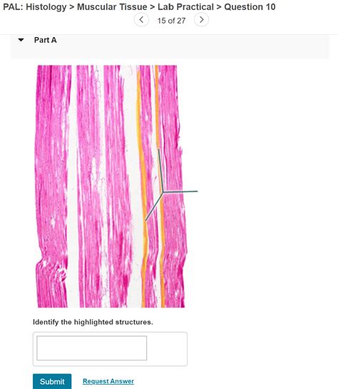
- Key Characteristics: Skeletal muscle is characterized by long, cylindrical, multinucleated fibers arranged in parallel bundles. These fibers exhibit striations, a hallmark feature resulting from the organized arrangement of actin and myosin filaments within sarcomeres. The nuclei are peripherally located.
- Identification under the microscope: Look for the characteristic striations, the multinucleated fibers, and the peripheral nuclei. The fibers are generally quite large and easily identifiable. Consider the tissue's location—skeletal muscle is typically associated with bones and tendons.
2. Cardiac Muscle Tissue: The Heart's Engine
- Key Characteristics: Cardiac muscle is composed of branched, uninucleated cells interconnected by intercalated discs. These discs are specialized junctions that facilitate rapid communication and coordinated contraction between cells. Striations are also present, though less pronounced than in skeletal muscle.
- Identification under the microscope: The branched nature of the cells and the presence of intercalated discs are definitive identifiers. Note the single, centrally located nucleus in each cell. Look for a more irregular arrangement compared to the parallel bundles of skeletal muscle.
3. Smooth Muscle Tissue: The Involuntary Controller
- Key Characteristics: Smooth muscle cells are spindle-shaped, uninucleated, and lack striations. They are responsible for involuntary movements in the walls of internal organs, blood vessels, and other structures.
- Identification under the microscope: The spindle shape of the cells and the absence of striations are key characteristics. The nuclei are elongated and centrally located. Often, smooth muscle appears as sheets or layers of cells.
Deconstructing "Question 9": A Hypothetical Example
Let's assume "Question 9" presents a microscopic image of muscular tissue and asks for identification and justification. To effectively answer, follow these steps:
-
Assess the Magnification: Determine the magnification level of the provided image. This is crucial for proper interpretation of cellular structures. Higher magnification will reveal finer details.
-
Identify the Basic Cell Shape and Arrangement: Observe the overall shape and arrangement of the muscle cells. Are they long and cylindrical, branched, or spindle-shaped? Are they arranged in parallel bundles or in a more haphazard fashion?
-
Examine for Striations: Carefully scan the cells for striations. Their presence or absence is a key differentiating factor between striated (skeletal and cardiac) and smooth muscle.
-
Locate and Describe the Nuclei: Identify the number and location of nuclei within each cell. Are they multiple and peripheral (skeletal muscle), single and central (cardiac and smooth muscle), or elongated and centrally located (smooth muscle)?
-
Look for Specialized Structures: Look for the presence of intercalated discs in cardiac muscle, which are unique to this type.
-
Deduce the Muscle Type and Justify Your Answer: Based on your observations, determine the muscle type (skeletal, cardiac, or smooth). Write a concise yet detailed justification based on the specific histological features you have identified. For example: "This is skeletal muscle tissue because it exhibits clear striations, multinucleated fibers with peripherally located nuclei, and a parallel arrangement of fibers."
Beyond "Question 9": Advanced Considerations
The ability to identify muscle tissue types is fundamental. However, a deeper understanding encompasses several crucial aspects:
1. Muscle Fiber Organization: A Closer Look
While the basic organization (parallel bundles in skeletal muscle, branched network in cardiac muscle, sheets or layers in smooth muscle) is essential, understanding variations within these patterns enhances your expertise. Consider variations in fiber size, density, and the presence of connective tissue within the muscle.
2. Connective Tissue Associations: The Supporting Structure
Pay attention to the connective tissue surrounding muscle fibers and bundles. Epimysium, perimysium, and endomysium play crucial roles in organizing and supporting the muscle tissue. Their presence and characteristics can aid in identification.
3. Artifacts and Staining Variations: Interpreting Imperfections
Microscopic slides may exhibit artifacts due to processing, staining, or other factors. Understanding potential artifacts is crucial to avoiding misinterpretations. Variations in staining intensity can also affect the visibility of cellular components.
4. Pathological Changes: Recognizing Aberrations
Histological examination is vital in diagnosing muscle diseases. Understanding the microscopic changes associated with common muscle pathologies is a critical skill for advanced studies.
5. Clinical Relevance: Connecting Histology to Physiology and Pathology
The histological identification of muscle tissue is not an isolated skill. It directly connects to understanding the physiology and pathology of the muscular system. For example, recognizing changes in muscle fiber size or the presence of inflammatory cells can indicate muscle diseases.
Mastering Muscle Tissue Histology: Practical Tips
- Practice, practice, practice: The key to mastering muscle tissue identification is consistent practice with microscopic images and slides.
- Utilize online resources: Numerous online histology atlases and resources offer high-quality images and detailed descriptions of various muscle types.
- Collaborate with classmates: Studying and discussing histology with peers can enhance your understanding and learning.
- Engage with your instructor: Don't hesitate to ask questions and seek clarification from your instructor if you are encountering difficulties.
- Develop a systematic approach: Adopt a structured method for analyzing microscopic images, as outlined earlier.
By diligently applying these strategies and expanding your understanding beyond a simple answer to "Question 9," you will build a solid foundation in muscular tissue histology. This knowledge will serve you well in future studies, research, and clinical practice. Remember, histology is a visual science—the more you practice observing and analyzing microscopic images, the more confident and proficient you will become.
Latest Posts
Latest Posts
-
How To Read Literature Like A Professor Chapters
Apr 01, 2025
-
Summary Of The Gilded Six Bits
Apr 01, 2025
-
The Earliest Form Of Intraverbal Training Is
Apr 01, 2025
-
Guided Reading Activity Industrialization 1865 To 1901 Answers
Apr 01, 2025
-
Which Passage Is The Best Example Of Deductive Reasoning
Apr 01, 2025
Related Post
Thank you for visiting our website which covers about Pal Histology Muscular Tissue Lab Practical Question 9 . We hope the information provided has been useful to you. Feel free to contact us if you have any questions or need further assistance. See you next time and don't miss to bookmark.
