Pre Lab Exercise 20-2 Formed Elements
Onlines
Mar 14, 2025 · 6 min read
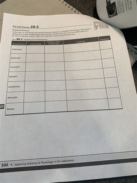
Table of Contents
Pre-Lab Exercise: Formed Elements of Blood (20-2) – A Comprehensive Guide
Understanding the formed elements of blood is crucial for comprehending the intricacies of human physiology and pathology. This pre-lab exercise delves into the fascinating world of erythrocytes, leukocytes, and thrombocytes, exploring their structure, function, and clinical significance. This guide provides a thorough overview, exceeding the typical pre-lab requirements, equipping you with a comprehensive understanding to excel in your laboratory session and beyond.
I. Introduction: The Composition of Blood
Blood, the lifeblood of the body, is a complex fluid connective tissue. It's not just a homogenous liquid; rather, it's a suspension of various cellular components – the formed elements – within a liquid matrix called plasma. This pre-lab exercise focuses specifically on the formed elements, which constitute a significant portion of blood's functionality. We will explore each element in detail, examining their microscopic features, physiological roles, and the implications of their abnormalities.
Keywords: Formed elements, blood, erythrocytes, leukocytes, thrombocytes, plasma, hematology, physiology, pathology, microscopy, clinical significance.
II. Erythrocytes: The Oxygen Carriers
Erythrocytes, commonly known as red blood cells (RBCs), are the most abundant formed elements. Their primary function is oxygen transport from the lungs to the body's tissues and carbon dioxide transport from the tissues back to the lungs.
A. Structure and Function of Erythrocytes:
- Biconcave shape: This unique shape maximizes surface area for efficient gas exchange. The flexible membrane allows RBCs to navigate through even the narrowest capillaries.
- Hemoglobin: This iron-containing protein is the key player in oxygen transport. Each hemoglobin molecule can bind to four oxygen molecules. The structure and function of hemoglobin are vital for understanding oxygen saturation levels and oxygen-carrying capacity.
- Anucleate: Mature RBCs lack a nucleus and other organelles, maximizing space for hemoglobin. This also means they have a limited lifespan and are constantly being replaced.
- Metabolic pathways: Despite lacking organelles, RBCs possess crucial metabolic pathways to maintain their function, primarily anaerobic glycolysis for energy production.
B. Clinical Significance of Erythrocyte Abnormalities:
- Anemia: Characterized by a reduced number of RBCs or decreased hemoglobin levels, leading to insufficient oxygen transport. Various types of anemia exist, each with distinct causes and manifestations. Understanding the different types is crucial for appropriate diagnosis and treatment.
- Polycythemia: This condition involves an abnormally high number of RBCs, potentially increasing blood viscosity and leading to cardiovascular complications.
- Sickle cell anemia: A genetic disorder causing abnormal hemoglobin, resulting in misshapen RBCs that can obstruct blood flow. This leads to chronic pain, organ damage, and other serious health problems.
- Thalassemia: A group of inherited blood disorders characterized by reduced or absent globin chain synthesis, impacting hemoglobin production and resulting in anemia.
Keywords: Red blood cells, hemoglobin, oxygen transport, carbon dioxide transport, biconcave shape, anemia, polycythemia, sickle cell anemia, thalassemia, hematocrit, mean corpuscular volume (MCV).
III. Leukocytes: The Immune Defenders
Leukocytes, also known as white blood cells (WBCs), are essential components of the body's immune system. They defend against infection and disease through various mechanisms. Unlike erythrocytes, leukocytes possess a nucleus and other organelles. They are classified into two main groups: granulocytes and agranulocytes.
A. Granulocytes:
- Neutrophils: The most abundant type of WBC, they are phagocytic cells, engulfing and destroying bacteria and other pathogens. Their multi-lobed nucleus is a distinguishing characteristic. Increased neutrophil counts often indicate bacterial infection.
- Eosinophils: These cells play a crucial role in combating parasitic infections and allergic reactions. Their characteristic bilobed nucleus and large granules are easily identifiable under a microscope.
- Basophils: The least abundant granulocytes, basophils release histamine and heparin, contributing to inflammatory responses and anticoagulation. Their dark-staining granules often obscure the nucleus.
B. Agranulocytes:
- Lymphocytes: These cells are key players in adaptive immunity. They include B lymphocytes (producing antibodies) and T lymphocytes (involved in cell-mediated immunity). Their large, round nucleus is a prominent feature. Variations in lymphocyte counts can indicate viral infections or immune disorders.
- Monocytes: These are the largest WBCs, acting as phagocytes and antigen-presenting cells, bridging the innate and adaptive immune systems. They differentiate into macrophages in tissues.
C. Clinical Significance of Leukocyte Abnormalities:
- Leukocytosis: An abnormally high WBC count, often indicating infection, inflammation, or certain blood cancers.
- Leukopenia: An abnormally low WBC count, increasing susceptibility to infections.
- Leukemia: A group of cancers affecting the blood-forming tissues, resulting in uncontrolled production of abnormal WBCs. Different types of leukemia exist, with varying prognoses and treatments.
- Infectious mononucleosis ("mono"): A viral infection causing an increase in lymphocytes, often associated with fatigue and swollen lymph nodes.
Keywords: White blood cells, granulocytes, agranulocytes, neutrophils, eosinophils, basophils, lymphocytes, monocytes, phagocytosis, adaptive immunity, innate immunity, leukocytosis, leukopenia, leukemia, infectious mononucleosis, differential white blood cell count.
IV. Thrombocytes: The Clotting Agents
Thrombocytes, also known as platelets, are small, irregular-shaped cell fragments essential for blood clotting (hemostasis). They lack a nucleus but contain various granules crucial for their function.
A. Structure and Function of Thrombocytes:
- Cell fragments: Platelets are derived from megakaryocytes in bone marrow.
- Granules: These contain factors involved in clot formation, including clotting proteins and growth factors.
- Activation and aggregation: Upon vascular injury, platelets adhere to the exposed collagen, becoming activated and aggregating to form a platelet plug. This plug is crucial in initiating the coagulation cascade.
- Coagulation factors: Platelets release various factors that trigger the coagulation cascade, a complex series of enzymatic reactions leading to fibrin formation and clot stabilization.
B. Clinical Significance of Thrombocyte Abnormalities:
- Thrombocytopenia: A low platelet count, increasing the risk of bleeding. Various conditions, including autoimmune diseases and certain medications, can cause thrombocytopenia.
- Thrombocytosis: An elevated platelet count, increasing the risk of thrombosis (blood clot formation), potentially leading to stroke or heart attack.
- Inherited bleeding disorders: Genetic defects affecting platelet function or coagulation factors can result in excessive bleeding. Examples include von Willebrand disease and hemophilia.
Keywords: Platelets, thrombocytes, hemostasis, blood clotting, coagulation, platelet plug, thrombocytopenia, thrombocytosis, von Willebrand disease, hemophilia, coagulation cascade, fibrin.
V. Laboratory Techniques for Studying Formed Elements
This section briefly outlines the common laboratory techniques used to analyze blood's formed elements:
- Complete Blood Count (CBC): A comprehensive blood test providing information on RBC count, WBC count, platelet count, hemoglobin levels, hematocrit, and more. A CBC is a fundamental diagnostic tool in hematology.
- Blood smear microscopy: A stained blood smear allows for visual examination of the formed elements under a microscope, enabling identification of different cell types and assessment of their morphology. This technique is crucial for diagnosing various hematological conditions.
- Differential white blood cell count: This specific analysis quantifies the proportions of different types of WBCs in a blood sample. Deviations from normal ranges can provide insights into the underlying cause of abnormal WBC counts.
- Platelet function tests: These tests assess platelet function and ability to form clots, helping to diagnose bleeding disorders.
VI. Conclusion: The Interconnectedness of Formed Elements
The formed elements of blood are not isolated entities; they work together in a complex and coordinated manner to maintain homeostasis. Understanding their individual roles and their interactions is crucial for interpreting laboratory results and comprehending the pathophysiology of various diseases. This pre-lab exercise provides a solid foundation for your upcoming laboratory session and your future studies in hematology and related fields. Remember to actively engage in the lab activities, paying close attention to the microscopic features of each formed element. Thorough observation and careful interpretation of your findings are key to mastering this important topic. The more you understand the individual elements and their interactions, the better you'll grasp the overall picture of blood’s vital function in maintaining health.
Latest Posts
Latest Posts
-
Symbolisms In A Good Man Is Hard To Find
Mar 14, 2025
-
Sample Recommendation Letter For National Junior Honor Society
Mar 14, 2025
-
Unit 8 Formative Assessment Common Core Algebra 1 Answer Key
Mar 14, 2025
-
The Accompanying Diagram Represents The Market For Violins
Mar 14, 2025
-
Copy Pq To The Line With An Endpoint At R
Mar 14, 2025
Related Post
Thank you for visiting our website which covers about Pre Lab Exercise 20-2 Formed Elements . We hope the information provided has been useful to you. Feel free to contact us if you have any questions or need further assistance. See you next time and don't miss to bookmark.
