Pre-lab Video Coaching Activity Stretch Reflexes
Onlines
Mar 26, 2025 · 7 min read
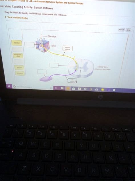
Table of Contents
Pre-Lab Video Coaching Activity: Stretch Reflexes
Understanding the stretch reflex is fundamental to grasping the complexities of the human nervous system. This pre-lab activity, utilizing video coaching, offers a unique opportunity to delve into the intricacies of this crucial neurological response before even stepping into the laboratory. This comprehensive guide will not only walk you through the theoretical underpinnings of the stretch reflex but also provide practical advice on how to effectively utilize video coaching to maximize your learning experience.
Understanding the Stretch Reflex: A Deep Dive
The stretch reflex, also known as the myotatic reflex, is a monosynaptic reflex arc that provides a rapid, involuntary response to muscle stretching. This seemingly simple reflex is incredibly important for maintaining posture, balance, and coordinating movement. Let's break down the key components:
The Players: Muscles, Sensory Neurons, and Motor Neurons
-
Muscle Spindles: These specialized sensory receptors, located within the muscle belly, are crucial for detecting changes in muscle length and rate of change of length. They're like tiny "stretch detectors" constantly monitoring the muscle's state. When a muscle is stretched, the muscle spindles are also stretched, activating sensory neurons.
-
Sensory Neurons (Ia afferents): These neurons transmit the sensory information from the muscle spindles to the spinal cord. They're the communication line relaying the "stretch detected" message.
-
Spinal Cord: The spinal cord acts as the central processing unit for this reflex. The sensory information from the Ia afferents synapses directly with the motor neurons.
-
Motor Neurons (α-motor neurons): These neurons receive the signal from the sensory neurons and transmit it to the muscle fibers causing them to contract. This is the command that triggers the muscle to counteract the stretch.
-
Muscle Fibers (Extrafusal fibers): These are the main muscle fibers responsible for producing movement. They receive the signal from the motor neurons and contract, leading to the reflexive muscle shortening.
The Pathway: A Monosynaptic Marvel
The simplicity and speed of the stretch reflex are due to its monosynaptic nature. This means that the sensory neuron directly synapses with the motor neuron in the spinal cord, without any interneurons involved. This direct connection ensures a rapid response—minimal delay between stimulus and response. This is critical for maintaining balance and stability. A delay would be catastrophic for quick adjustments needed for maintaining posture.
Reciprocal Inhibition: The Antagonist's Role
While the agonist muscle (the muscle that's stretched) contracts, its antagonist muscle (the muscle that opposes the agonist's action) simultaneously relaxes. This coordinated action is achieved through reciprocal inhibition. Interneurons within the spinal cord inhibit the motor neurons of the antagonist muscle, ensuring smooth, coordinated movement. Without reciprocal inhibition, the stretch reflex would be much less efficient and could result in jerky movements.
Clinical Significance: Assessing Neurological Function
The stretch reflex is routinely assessed during neurological examinations. The presence or absence, as well as the intensity, of the reflex provides valuable information about the integrity of the nervous system. Abnormal reflex responses can indicate damage to the peripheral nerves, spinal cord, or brain. Commonly tested reflexes include the patellar (knee-jerk) reflex, the Achilles (ankle-jerk) reflex, and the biceps reflex.
Pre-Lab Video Coaching: Maximizing Your Learning
Video coaching offers a powerful tool for pre-lab preparation. By watching and analyzing videos demonstrating the stretch reflex, you can gain a deeper understanding of the process before conducting the actual experiment. Here's how to effectively utilize video coaching:
Selecting High-Quality Videos
Look for videos that clearly demonstrate the following:
- Stimulus and Response: The video should clearly show the stimulus (e.g., tapping the patellar tendon) and the resulting muscle contraction.
- Muscle Anatomy: The video should ideally highlight the relevant muscles involved, including both the agonist and antagonist muscles.
- Neurological Pathway: Ideally, the video should provide a clear visual representation of the neurological pathway involved in the stretch reflex.
Active Viewing: Engaging with the Material
Don't passively watch the video. Instead, actively engage with the material by:
- Taking Notes: Jot down key observations, including the timing of the response, the amplitude of the muscle contraction, and any other relevant details.
- Drawing Diagrams: Create diagrams to illustrate the neurological pathway and the muscles involved. This will reinforce your understanding of the anatomical and physiological components.
- Formulating Questions: As you watch the video, formulate questions that you can address during the lab session. This is an effective way to anticipate challenges and plan for efficient data collection.
Utilizing Slow-Motion and Replay: Identifying Key Moments
Many videos offer the ability to slow down the playback speed. Use this feature to carefully observe the sequence of events in the stretch reflex. Replaying key moments can help you identify subtle details that you might miss during normal playback.
Peer Learning and Discussion: Sharing Insights
If possible, watch the videos with a study partner or in a small group. Discussing your observations and insights with others can deepen your understanding and help you identify any gaps in your knowledge. This collaborative approach will improve comprehension and can help to highlight specific areas for focus.
Connecting Theory to Practice: Preparing for the Lab
Before the lab session, make sure you can thoroughly describe the following:
- The components of the stretch reflex arc: Be able to clearly identify the sensory receptors, sensory neurons, motor neurons, and the role of the spinal cord.
- The monosynaptic nature of the reflex: Understand why this direct pathway is crucial for the rapid response.
- Reciprocal inhibition: Explain how the antagonist muscle is inhibited during the reflex.
- Clinical applications: Understand how the stretch reflex is used to assess neurological function.
By engaging in this thorough pre-lab video coaching, you will be fully prepared to perform and interpret the laboratory experiment effectively.
Further Exploration: Enhancing Understanding
Beyond the basic stretch reflex, there are several related concepts that are worth exploring to deepen your understanding:
Gamma Motor Neurons and Muscle Tone: Fine-Tuning the Reflex
Gamma motor neurons innervate the intrafusal fibers within the muscle spindles. These neurons regulate the sensitivity of the muscle spindles, allowing the body to adjust the stretch reflex based on the current activity level and needs of the body. This control allows for fine adjustments and maintain muscle tone. Understanding this adds complexity and detail to the simple model of the reflex arc.
Jendrassik Maneuver: Enhancing Reflex Responsiveness
The Jendrassik maneuver is a technique used to enhance the reflex response. This is achieved by distracting the subject by asking them to interlock their fingers and pull strongly. The underlying mechanisms are still not completely understood, but it is thought to inhibit the descending inhibitory pathways from the brain, allowing a more pronounced reflex response.
Pathological Reflexes: Indications of Neurological Dysfunction
Variations or absences in reflexes can signify underlying neurological conditions. Understanding these pathological reflexes, and their associated conditions, is critical for clinical practice. Examples include hyporeflexia (diminished reflexes), hyperreflexia (exaggerated reflexes), and clonus (rhythmic oscillations). This knowledge will further enhance your understanding of the role of the stretch reflex in overall neurological function.
The Role of Higher Brain Centers: Modulation and Control
While the stretch reflex is fundamentally a spinal reflex, higher brain centers, such as the brainstem and cerebellum, also play a role in modulating and controlling its response. This modulation allows for greater flexibility and adaptability in movement. This understanding adds another level of complexity, illustrating that reflexes are not simply fixed, hardwired responses, but are subject to ongoing regulatory influence.
Conclusion: A Powerful Tool for Learning
Pre-lab video coaching, when used effectively, is a powerful tool for enhancing your understanding of complex physiological processes like the stretch reflex. By actively engaging with the video material, preparing questions, and discussing your observations, you'll gain a solid foundation before entering the lab. This will significantly improve your learning outcomes, strengthen your practical skills, and provide a deeper appreciation of the fascinating complexities of the human nervous system. Remember to always connect the theoretical knowledge with the practical application and to utilize various learning techniques for effective knowledge retention.
Latest Posts
Latest Posts
-
Choose All Functions Typically Carried Out By Membrane Proteins
Mar 29, 2025
-
Table 1 Rate Of Diffusion In Corn Syrup
Mar 29, 2025
-
Comparative Anatomy Of The Domestic Chicken
Mar 29, 2025
-
Stone And Brick Are Substitutes In Home Construction
Mar 29, 2025
-
Damage To The Circled Area May Cause What Symptoms
Mar 29, 2025
Related Post
Thank you for visiting our website which covers about Pre-lab Video Coaching Activity Stretch Reflexes . We hope the information provided has been useful to you. Feel free to contact us if you have any questions or need further assistance. See you next time and don't miss to bookmark.
