Put The Following Mitosis And Cytokinesis Images In Order
Onlines
Mar 17, 2025 · 5 min read
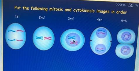
Table of Contents
Putting Mitosis and Cytokinesis Images in Order: A Comprehensive Guide
Understanding the phases of mitosis and cytokinesis is crucial for comprehending cell division and the fundamental processes of life. Visual aids, such as images, are invaluable tools for learning these complex stages. However, interpreting these images and placing them in the correct sequential order can be challenging. This article serves as a comprehensive guide to help you accurately sequence images depicting the stages of mitosis and cytokinesis, building a strong foundation in cell biology.
Understanding Mitosis and Cytokinesis: A Quick Recap
Before we delve into ordering images, let's briefly review the key stages of mitosis and cytokinesis. Mitosis is the process of nuclear division, resulting in two genetically identical daughter nuclei. Cytokinesis is the division of the cytoplasm, producing two separate daughter cells. These processes work in concert to ensure accurate cell duplication.
Mitosis is typically divided into several phases:
- Prophase: Chromosomes condense and become visible, the nuclear envelope breaks down, and the mitotic spindle begins to form.
- Prometaphase: The mitotic spindle fibers attach to the chromosomes at their kinetochores.
- Metaphase: Chromosomes align at the metaphase plate (the equator of the cell).
- Anaphase: Sister chromatids separate and move towards opposite poles of the cell.
- Telophase: Chromosomes arrive at the poles, the nuclear envelope reforms, and chromosomes decondense.
Cytokinesis, the division of the cytoplasm, overlaps with the later stages of mitosis, typically beginning in anaphase and concluding after telophase. The process differs slightly between plant and animal cells.
Identifying Key Features in Mitosis and Cytokinesis Images
Successfully ordering images requires a keen eye for detail. Here's a breakdown of the key visual features to look for in each stage:
1. Chromosome Condensation: Look for the progressive condensation of chromosomes. In early stages (prophase), they appear as long, thin threads. As the process advances, they become shorter, thicker, and more easily discernible.
2. Nuclear Envelope: The presence or absence of the nuclear envelope is a crucial indicator. It's intact in interphase and early prophase, but breaks down during prometaphase and reappears in telophase.
3. Mitotic Spindle: Observe the formation and function of the mitotic spindle. In prophase and prometaphase, it starts forming, and in metaphase, it's fully developed, with chromosomes aligned at the metaphase plate. In anaphase, it facilitates the separation of sister chromatids.
4. Chromosome Alignment: In metaphase, chromosomes are precisely aligned at the metaphase plate. This is a defining characteristic of this stage.
5. Sister Chromatid Separation: The separation of sister chromatids is a hallmark of anaphase. Look for individual chromosomes moving towards opposite poles.
6. Cleavage Furrow (Animal Cells) or Cell Plate (Plant Cells): Cytokinesis is evident by the formation of a cleavage furrow (in animal cells) which gradually pinches the cell in two, or a cell plate (in plant cells), which grows inward from the cell's center.
7. Formation of Two Nuclei: The appearance of two distinct nuclei is a clear indication of telophase.
A Step-by-Step Approach to Ordering Mitosis and Cytokinesis Images
To effectively sequence the images, follow these steps:
-
Identify Interphase (if present): If you have an image showing a cell before mitosis, it will be in interphase. Chromosomes are not condensed and the nuclear envelope is intact.
-
Locate Prophase: Look for an image showing condensed chromosomes, but with the nuclear envelope still present or starting to break down.
-
Find Prometaphase: The nuclear envelope should be gone, and spindle fibers should be attaching to the chromosomes.
-
Identify Metaphase: The defining feature is the alignment of chromosomes at the metaphase plate.
-
Spot Anaphase: Look for the separation of sister chromatids and their movement towards opposite poles.
-
Recognize Telophase: The chromosomes have reached the poles, the nuclear envelope reforms, and chromosomes begin to decondense.
-
Observe Cytokinesis: Look for either a cleavage furrow (animal cells) or a cell plate (plant cells) indicating the division of the cytoplasm.
Common Mistakes to Avoid
When ordering mitosis and cytokinesis images, several common mistakes can occur:
-
Confusing prophase and metaphase: Pay close attention to chromosome alignment. In prophase, chromosomes are not yet aligned at the metaphase plate.
-
Misinterpreting anaphase and telophase: Sister chromatid separation is unique to anaphase. In telophase, chromosomes have already reached the poles.
-
Ignoring cytokinesis: Don't overlook the signs of cytokinesis, as it's an essential part of cell division.
-
Assuming all images are from the same cell type: Remember that cytokinesis differs between animal and plant cells.
Advanced Considerations: Variations and Exceptions
While the above steps provide a general framework, it's crucial to remember that cell division is a dynamic process. Slight variations can occur depending on the cell type and experimental conditions. Some images might show intermediate stages, making the exact identification challenging. In these instances, rely on the overall progression of events – chromosome condensation, spindle formation, chromatid separation, and nuclear envelope reformation.
Enhancing your Understanding Through Practice
The best way to master image sequencing is through practice. Search online for various images of mitosis and cytokinesis and try to arrange them in the correct order. Compare your sequence to the descriptions and visuals provided in textbooks or online resources. This repeated practice will enhance your understanding and sharpen your observational skills. Consider also looking at videos of mitosis and cytokinesis; this dynamic representation can help considerably in visual understanding.
Conclusion
Ordering images of mitosis and cytokinesis requires careful observation of key features and an understanding of the sequential events in cell division. By following the steps outlined in this article and practicing regularly, you can develop the skills to confidently sequence images and strengthen your knowledge of this fundamental biological process. This enhanced understanding will prove invaluable in your broader studies of cell biology and related fields. Remember, consistent practice is key to mastering this skill and reinforcing your grasp of mitosis and cytokinesis.
Latest Posts
Latest Posts
-
Select The Correct Answer From Each Drop Down Menu
Mar 17, 2025
-
Christian High School Equivalency Exam Answers
Mar 17, 2025
-
Introduction To Health Assessment 3 0 Test
Mar 17, 2025
-
Lord Of The Flies Student Workbook Answers Pdf
Mar 17, 2025
-
In Time Of The Butterflies Quotes
Mar 17, 2025
Related Post
Thank you for visiting our website which covers about Put The Following Mitosis And Cytokinesis Images In Order . We hope the information provided has been useful to you. Feel free to contact us if you have any questions or need further assistance. See you next time and don't miss to bookmark.
