Record The Relevant Values Of Your Microscope
Onlines
Mar 31, 2025 · 6 min read

Table of Contents
Recording the Relevant Values of Your Microscope: A Comprehensive Guide
Microscopy, a cornerstone of scientific research and various industrial applications, demands meticulous record-keeping. Failing to accurately document the settings and parameters used during observation can lead to irreproducible results, compromised data integrity, and ultimately, flawed conclusions. This comprehensive guide details the critical values to record for different types of microscopes, emphasizing best practices for maintaining accurate and readily accessible records.
Why Accurate Microscope Record Keeping is Crucial
The importance of diligent record-keeping in microscopy cannot be overstated. Consider these key reasons:
Reproducibility of Results: Scientific research relies heavily on the ability to replicate experiments. Without detailed records of microscope settings, repeating an experiment to verify results becomes virtually impossible. This is particularly important in fields like pathology, materials science, and nanotechnology where minute details can significantly impact interpretations.
Data Integrity and Accuracy: Accurate records ensure data integrity by providing a complete and verifiable account of the experimental process. This prevents ambiguity and errors in data analysis and reporting. Omitting crucial details can lead to misinterpretations and invalidate research findings.
Troubleshooting and Maintenance: Detailed logs can be invaluable when troubleshooting issues with the microscope. By reviewing past records, you can identify potential sources of error or malfunction. This proactive approach reduces downtime and ensures the longevity of the instrument.
Collaboration and Communication: When collaborating with colleagues or sharing data, comprehensive records are essential for clear communication. They provide a shared understanding of the experimental setup and parameters used, facilitating efficient collaboration and the avoidance of misunderstandings.
Compliance and Regulatory Requirements: Many industries and research institutions have specific guidelines and regulations regarding data management and record-keeping. Detailed microscope records are often crucial for compliance purposes and ensuring the validity of research findings for publication or regulatory submissions.
What Values to Record: A Microscope-Specific Approach
The specific values you need to record depend heavily on the type of microscope being used. Let's examine this for common microscope types:
Optical Microscopes (Brightfield, Darkfield, Phase Contrast, Fluorescence):
-
**Microscope Model and Serial Number: This uniquely identifies your instrument.
-
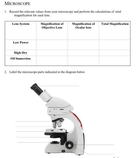
**Objective Lens: Record the magnification (e.g., 4x, 10x, 40x, 100x) and numerical aperture (NA) of each objective used. The NA indicates the lens's ability to gather light and resolve fine details.
-
**Eyepiece Magnification: Note the magnification of the eyepieces. Total magnification is calculated by multiplying the objective and eyepiece magnifications.
-
**Immersion Medium (if applicable): If using an oil immersion objective (typically 100x), specify the type of immersion oil used (e.g., type A, type B).
-
**Illumination Source: Specify the type of light source (e.g., halogen, LED, mercury arc lamp for fluorescence) and its intensity settings. For fluorescence microscopy, record the excitation and emission wavelengths used.
-
**Condenser Settings: Record the condenser aperture diaphragm setting (often a numerical scale or marked positions). This controls the amount of light entering the specimen.
-
**Focus Settings: Although generally not recorded precisely, note the approximate focal plane used (e.g., coarse focus, fine focus). For high-precision work, record precise measurements of the fine focus adjustment.
-
**Filters: Record the use of any filters (e.g., neutral density, excitation, emission filters for fluorescence) and their specifications.
-
**Specimen Details: This is absolutely crucial. Include:
- Specimen Identification: A unique identifier for your specimen (e.g., slide number, sample code).
- Specimen Preparation: Describe the preparation method used (e.g., staining technique, embedding medium, section thickness).
- Mounting Medium: Specify the mounting medium used (if applicable).
- Date and Time of Observation: Record the exact date and time of observation.
-
**Image Capture Settings (if applicable): If capturing images, record the camera settings used, including:
- Exposure Time: The duration of light exposure.
- Gain: The amplification of the signal.
- Resolution: The image resolution (e.g., pixels).
- Software Used: Specify the image acquisition software used.
Electron Microscopes (TEM, SEM):
Electron microscopy requires even more detailed record-keeping due to the complexities of sample preparation and instrument operation. In addition to the general points above (date, time, sample identification), you should record:
-
**Microscope Model and Serial Number: Critical for traceability.
-
**Accelerating Voltage: The voltage used to accelerate the electrons (kV).
-
**Beam Current: The intensity of the electron beam.
-
**Working Distance: The distance between the objective lens and the specimen.
-
**Aperture Size: The size of the aperture used to control the electron beam.
-
**Magnification: Record the magnification used.
-
**Detector Type and Settings: Specify the type of detector used (e.g., secondary electron detector, backscattered electron detector) and its settings (e.g., gain, offset).
-
**Specimen Preparation: Provide detailed information on the specimen preparation method, including:
- Fixation: The method used to preserve the specimen.
- Dehydration: The method used to remove water from the specimen.
- Embedding: The method used to embed the specimen in a resin.
- Sectioning: The method used to cut thin sections of the specimen (for TEM).
- Coating: The type of coating applied (if applicable, for SEM).
-
**Image Capture Settings: Similar to optical microscopy, record the image capture parameters, including resolution, exposure time, and software used. For TEM, note the type of imaging mode used (e.g., bright field, dark field).
Confocal Microscopes:
Confocal microscopes offer advanced imaging capabilities requiring specific record-keeping:
-
**Microscope Model and Serial Number: Essential for traceability.
-
**Laser Wavelengths: Record the wavelengths of the lasers used for excitation.
-
**Pinhole Size: Record the size of the pinhole, which affects the resolution and depth of field.
-
**Scan Speed: The speed at which the laser scans the specimen.
-
**Gain and Offset: The settings used to adjust the detector sensitivity.
-
**Z-Stack Parameters: If acquiring a Z-stack (a series of images at different focal planes), record the step size and the number of images acquired.
-
**Software Used: Specify the image acquisition and processing software used.
Best Practices for Record Keeping
Beyond the specific values, adopting consistent best practices enhances the quality and usefulness of your microscope records:
-
**Use a Standardized Format: Create a standardized template or spreadsheet to ensure consistency in recording information. This facilitates data analysis and comparison.
-
**Utilize a Laboratory Information Management System (LIMS): LIMS software provides a structured approach to managing laboratory data, including microscope records.
-
**Maintain a Detailed Laboratory Notebook: Combine digital records with a traditional laboratory notebook to provide a comprehensive and readily accessible record.
-
**Use Clear and Concise Language: Avoid ambiguity and jargon. Use precise and descriptive language to accurately convey information.
-
**Regularly Back Up Your Data: Digital records should be backed up regularly to prevent data loss.
-
**Store Records Securely: Ensure that your microscope records are stored securely and are accessible only to authorized personnel.
-
**Use a version control system for image data: Employ a system (like Git LFS) that tracks changes to large files such as images, permitting restoration to prior versions if needed.
Conclusion
Recording relevant values from your microscope is not merely a formality; it's a fundamental aspect of ensuring the reproducibility, integrity, and validity of your microscopy work. By following the guidelines and best practices outlined in this comprehensive guide, you can establish a robust system for managing your microscope records, contributing to the accuracy and reliability of your research and applications. The time invested in meticulous record-keeping pays significant dividends in the long run, safeguarding your data and furthering your scientific endeavors.
Latest Posts
Latest Posts
-
Night Chapter 5 Questions And Answers Pdf
Apr 02, 2025
-
Carter Racing Case Study Solution Pdf
Apr 02, 2025
-
A Good Behavioral Definition Of A Behavior Involves
Apr 02, 2025
-
Completa Estas Oraciones Con Las Preposiciones Por O Para
Apr 02, 2025
-
Color By Number Natural Selection Answers
Apr 02, 2025
Related Post
Thank you for visiting our website which covers about Record The Relevant Values Of Your Microscope . We hope the information provided has been useful to you. Feel free to contact us if you have any questions or need further assistance. See you next time and don't miss to bookmark.
