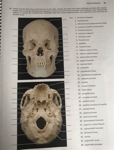
The Axial Skeleton Review Sheet 8
Onlines
Apr 04, 2025 · 7 min read

Table of Contents
The Axial Skeleton: A Comprehensive Review (Sheet 8)
This comprehensive review sheet delves into the intricacies of the axial skeleton, providing a detailed overview ideal for students, medical professionals, or anyone seeking a deeper understanding of this crucial part of the human anatomy. We will cover key features, functions, and clinical considerations, ensuring a thorough understanding. This guide surpasses a simple "sheet 8" by providing in-depth explanations and contextual information, making learning engaging and effective.
I. Introduction to the Axial Skeleton
The axial skeleton forms the central axis of the body, providing structural support and protection for vital organs. Unlike the appendicular skeleton (limbs and girdles), it's primarily responsible for maintaining posture, enabling movement of the head and trunk, and safeguarding the nervous system and other critical structures. Key components include:
- The Skull: Protecting the brain and housing sensory organs.
- The Vertebral Column: Providing support for the body, protecting the spinal cord, and enabling flexibility.
- The Thoracic Cage: Protecting the heart and lungs and playing a crucial role in respiration.
This review focuses on each of these components in detail.
II. The Skull: A Protective Fortress
The skull, or cranium, is a complex structure composed of numerous bones fused together to form a rigid protective shell. We'll break it down into two main parts:
A. Neurocranium (Braincase):
- Frontal Bone: Forms the forehead and part of the anterior cranial fossa. Key features include the supraorbital ridges and the frontal sinuses. Consider its role in facial expression and protection of the frontal lobes of the brain.
- Parietal Bones (2): Form the majority of the superior and lateral aspects of the cranium. Note their articulation with other cranial bones, including the frontal, occipital, temporal, and sphenoid bones.
- Occipital Bone: Forms the posterior and inferior aspects of the cranium. Key features include the foramen magnum (passage for the spinal cord), occipital condyles (articulation with the atlas), and the external occipital protuberance. Consider its role in supporting the head and its connection to the vertebral column.
- Temporal Bones (2): Located on the lateral sides of the skull, they house the organs of hearing and balance. Key features include the zygomatic process (forms part of the cheekbone), mandibular fossa (articulation with the mandible), external acoustic meatus (ear canal), mastoid process, and styloid process.
- Sphenoid Bone: A complex, bat-shaped bone located at the base of the skull. It articulates with numerous other bones and contains important foramina (openings) for cranial nerves and blood vessels. Note its central location and its role in cranial stability and neurovascular pathways.
- Ethmoid Bone: Located anterior to the sphenoid bone, it contributes to the formation of the nasal cavity and orbits. Key features include the cribriform plate (passage for olfactory nerves) and the superior and middle nasal conchae.
B. Viscerocranium (Facial Bones):
- Maxillae (2): Form the upper jaw and part of the hard palate. Note their role in chewing, facial structure, and the housing of the maxillary sinuses.
- Mandible: Forms the lower jaw, the only movable bone of the skull. Consider its function in mastication (chewing) and speech.
- Zygomatic Bones (2): Form the cheekbones. Note their contribution to facial structure and protection of the underlying soft tissues.
- Nasal Bones (2): Form the bridge of the nose. Consider their role in shaping the nasal cavity.
- Lacrimal Bones (2): Small bones forming part of the medial wall of the orbit. Note their involvement in tear drainage.
- Vomer: Forms part of the nasal septum.
- Palatine Bones (2): Contribute to the hard palate and the posterior portion of the nasal cavity.
- Inferior Nasal Conchae (2): Scroll-like bones within the nasal cavity that increase surface area for air filtration and warming.
III. The Vertebral Column: The Body's Central Support
The vertebral column, or spine, is a flexible column of 33 vertebrae, divided into five regions:
- Cervical Vertebrae (7): The vertebrae of the neck. C1 (atlas) and C2 (axis) are unique in their structure and function, enabling head rotation and nodding. Note the transverse foramina (passage for vertebral arteries) in most cervical vertebrae.
- Thoracic Vertebrae (12): The vertebrae of the thoracic region, articulating with the ribs. Note the costal facets (articulation points for ribs).
- Lumbar Vertebrae (5): The largest and strongest vertebrae, supporting most of the body's weight. Note their robust structure.
- Sacral Vertebrae (5): Fused into the sacrum, forming the posterior wall of the pelvis.
- Coccygeal Vertebrae (4): Fused into the coccyx (tailbone).
Each vertebra consists of a body, vertebral arch, and various processes. Understanding these components is crucial for understanding vertebral function and potential pathologies. Consider the intervertebral discs, which act as shock absorbers between vertebrae. Note the curvature of the spine (cervical lordosis, thoracic kyphosis, lumbar lordosis, and sacral kyphosis). Abnormal curvatures are significant clinical considerations.
IV. The Thoracic Cage: Protecting Vital Organs
The thoracic cage, or rib cage, consists of:
- Sternum: The breastbone, comprising the manubrium, body, and xiphoid process.
- Ribs (12 pairs): True ribs (1-7) articulate directly with the sternum via costal cartilage. False ribs (8-10) articulate indirectly with the sternum through the costal cartilage of rib 7. Floating ribs (11-12) do not articulate with the sternum.
The thoracic cage protects the heart and lungs, and plays a vital role in respiration through its movement during inhalation and exhalation. Consider the articulations between ribs, vertebrae, and the sternum. Note the intercostal spaces between the ribs, containing intercostal muscles involved in respiration.
V. Clinical Considerations of the Axial Skeleton
Several clinical conditions can affect the axial skeleton:
- Fractures: Skull fractures (e.g., depressed fractures, basilar fractures), vertebral fractures (e.g., compression fractures, burst fractures), rib fractures. Note the varied severity and potential complications of each type of fracture.
- Spinal Stenosis: Narrowing of the spinal canal, causing compression of the spinal cord or nerve roots.
- Scoliosis: Lateral curvature of the spine.
- Kyphosis: Excessive thoracic curvature (hunchback).
- Lordosis: Excessive lumbar curvature (swayback).
- Osteoporosis: Weakening of the bones, increasing the risk of fractures.
- Spondylolysis and Spondylolisthesis: Stress fractures or slippage of vertebrae, often affecting the lumbar spine. Note the implications for back pain and potential neurological deficits.
- Cervical Spondylosis: Degenerative changes in the cervical spine, potentially causing neck pain and radiculopathy.
- Craniosynostosis: Premature fusion of cranial sutures, leading to abnormal skull shape.
VI. Developmental Considerations
The development of the axial skeleton is a complex process involving several stages:
- Intramembranous Ossification: Formation of certain skull bones directly from mesenchymal tissue.
- Endochondral Ossification: Formation of most bones of the axial skeleton from cartilage models.
- Vertebral Column Development: Formation of the vertebral column involves the segmentation of the somites and the development of the notochord. Note the clinical implications of developmental abnormalities.
- Rib Cage Development: The development of the rib cage involves the formation of the ribs from costal processes and their articulation with the sternum and vertebrae.
VII. Imaging Techniques and Examination
Various imaging techniques are employed for assessing the axial skeleton:
- X-rays: Provide basic images of bone structure, identifying fractures and other abnormalities.
- CT scans: Offer detailed cross-sectional images, useful for visualizing complex fractures and assessing soft tissues.
- MRI scans: Provide high-resolution images of soft tissues, useful for evaluating the spinal cord, intervertebral discs, and other soft tissue structures.
- Bone Densitometry: Measures bone mineral density, used for diagnosing osteoporosis.
VIII. Conclusion
The axial skeleton is a critical component of the human body, providing structural support, protection, and enabling essential functions. A comprehensive understanding of its anatomy, physiology, and clinical considerations is paramount for healthcare professionals and anyone seeking a deeper knowledge of human biology. This detailed review should provide a robust foundation for further study and clinical practice. Remember to consult reputable anatomical texts and resources for further in-depth learning. This review sheet aims to serve as a robust starting point for exploration. The complexity of the axial skeleton necessitates continued learning and exploration.
Latest Posts
Latest Posts
-
A Means To Guarantee Continuity In Career Development
Apr 05, 2025
-
2 1 The Nature Of Matter Answer Key
Apr 05, 2025
-
How Many Emails Were Sent Out Per Day In 2015
Apr 05, 2025
-
Unit 5 Progress Check Frq Ap Chemistry
Apr 05, 2025
-
Label A Label B Label C Label D
Apr 05, 2025
Related Post
Thank you for visiting our website which covers about The Axial Skeleton Review Sheet 8 . We hope the information provided has been useful to you. Feel free to contact us if you have any questions or need further assistance. See you next time and don't miss to bookmark.
