The Iliac Arteries Immediately Subdivide Into The
Onlines
Mar 06, 2025 · 6 min read
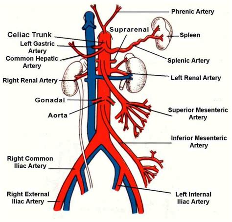
Table of Contents
The Iliac Arteries: Immediate Subdivisions and Clinical Significance
The iliac arteries represent a crucial segment of the lower abdominal arterial system, responsible for supplying oxygenated blood to the pelvis, buttocks, and lower limbs. Understanding their immediate subdivisions is paramount for clinicians, radiologists, and anyone involved in the diagnosis and treatment of vascular pathologies. This comprehensive article delves into the intricate anatomy of the iliac arteries, their branching patterns, clinical correlations, and potential implications for various medical conditions.
Anatomy of the Iliac Arteries: A Detailed Overview
The common iliac arteries arise from the bifurcation of the abdominal aorta, typically at the level of the fourth lumbar vertebra (L4). Each common iliac artery then further divides into two main branches: the internal iliac artery (hypogastric artery) and the external iliac artery. This bifurcation marks a critical anatomical landmark, often utilized during surgical procedures and radiological interventions.
1. Internal Iliac Artery (Hypogastric Artery): A Branching Network to the Pelvis
The internal iliac artery, a shorter and thicker vessel compared to its external counterpart, primarily supplies the pelvic organs and surrounding structures. Its branching pattern is highly variable, but several consistent branches are commonly identified:
-
Anterior Division: This division predominantly supplies the pelvic viscera and perineum. Key branches include:
- Umbilical artery: A remnant of the fetal circulation, its distal portion typically becomes obliterated, forming the medial umbilical ligament. However, the proximal portion persists and gives rise to the superior vesical artery, supplying the superior surface of the urinary bladder.
- Superior vesical artery: Supplies the superior part of the urinary bladder.
- Inferior vesical artery: Supplies the inferior part of the urinary bladder and the prostate (in males) or the vagina (in females).
- Obturator artery: Passes through the obturator canal to supply the medial compartment of the thigh. It's known for its occasional variations, with potential accessory obturator arteries arising from the inferior epigastric artery.
- Uterine artery (in females): Courses along the lateral aspect of the uterus and provides blood supply to the uterus and vagina. Its tortuous course is significant during gynecological procedures.
- Vaginal artery (in females): Supplies the vagina.
- Middle rectal artery: Supplies the rectum.
- Internal pudendal artery: Supplies the external genitalia and perineum. Its branches, such as the dorsal artery of the penis or clitoris, are crucial for erectile function.
- Inferior gluteal artery: Supplies the gluteal muscles.
-
Posterior Division: This division mainly supplies the gluteal muscles and the posterior pelvic wall. Notable branches include:
- Iliolumbar artery: Supplies the iliacus and quadratus lumborum muscles.
- Lateral sacral arteries: Supply the sacrum and the piriformis muscle.
- Superior gluteal artery: Supplies the gluteal muscles.
2. External Iliac Artery: The Pathway to the Lower Limb
The external iliac artery, continuing distally, is primarily responsible for supplying blood to the lower limb. It runs along the medial border of the psoas major muscle and enters the thigh by passing under the inguinal ligament, where it is then termed the femoral artery. Before this transition, it gives off one crucial branch:
- Inferior epigastric artery: Supplies the lower abdominal wall muscles. It is also notable for its potential role in supplying accessory obturator arteries.
Clinical Significance of Iliac Artery Subdivisions
Knowledge of the iliac arteries' anatomy and their branching patterns is essential in various clinical scenarios:
1. Atherosclerosis and Peripheral Artery Disease (PAD)
Atherosclerosis, the buildup of plaques within arterial walls, frequently affects the iliac arteries. This can lead to peripheral artery disease (PAD), characterized by reduced blood flow to the lower limbs. Symptoms include intermittent claudication (leg pain during exertion), rest pain, and potentially critical limb ischemia requiring amputation. Angiography, a radiological procedure involving the injection of contrast dye, is crucial for visualizing the iliac arteries and assessing the severity of atherosclerotic disease.
2. Iliac Artery Aneurysms
Iliac artery aneurysms, abnormal dilatations of the artery, can occur and pose a significant risk of rupture, leading to potentially fatal internal bleeding. Risk factors include smoking, hypertension, and hyperlipidemia. Diagnosis often involves imaging techniques such as ultrasound, CT angiography, or MRI angiography. Treatment options range from conservative management (close monitoring) to surgical intervention, including endovascular repair or open surgical repair.
3. Iliac Artery Embolization
Iliac artery embolization is a minimally invasive procedure used to treat various conditions. It involves the introduction of a catheter into the iliac artery to deliver therapeutic agents, such as embolic materials, to block blood flow to a specific area. This technique is utilized in the management of:
- Pelvic hemorrhage: Embolization can effectively control bleeding in cases of trauma or postpartum hemorrhage.
- Uterine fibroids: Embolization can reduce the size and blood supply to uterine fibroids, alleviating symptoms.
- Tumor embolization: In certain cancers involving the pelvic organs, embolization can reduce tumor growth and blood supply.
4. Iliac Vein Thrombosis
While not directly related to the arteries, the proximity of the iliac veins makes understanding their anatomy vital. Deep vein thrombosis (DVT) in the iliac veins can lead to significant complications, including pulmonary embolism (PE), a life-threatening condition. Diagnosis often involves ultrasound or venography.
5. Surgical Procedures
Detailed knowledge of iliac artery anatomy is crucial for various surgical procedures:
- Aortoiliac bypass: This procedure bypasses a blocked or narrowed section of the iliac artery to restore blood flow to the lower limb.
- Pelvic surgery: Surgeons must have a thorough understanding of the branching pattern of the internal iliac artery to avoid inadvertent injury during procedures involving pelvic organs.
- Vascular access: The iliac arteries can serve as access points for various vascular interventions, such as angiography and angioplasty.
Imaging Modalities for Iliac Artery Visualization
Several advanced imaging techniques are employed to visualize the iliac arteries and assess their condition:
- Ultrasound: A non-invasive method that uses high-frequency sound waves to create images of the arteries. It is commonly used for initial assessment and monitoring of vascular conditions.
- Computed Tomography Angiography (CTA): A sophisticated imaging technique that combines CT scanning with the injection of contrast dye to create detailed three-dimensional images of the arteries. It provides excellent visualization of the iliac arteries and their branches.
- Magnetic Resonance Angiography (MRA): A non-invasive technique using magnetic fields and radio waves to create images of the arteries. It is particularly useful for patients with contraindications to contrast dye.
- Conventional Angiography: A minimally invasive procedure that involves inserting a catheter into the artery and injecting contrast dye to directly visualize the blood vessels. It is often used for diagnostic and interventional purposes.
Conclusion
The iliac arteries, with their complex branching patterns, play a vital role in supplying blood to the pelvis and lower limbs. A thorough understanding of their anatomy and clinical significance is crucial for healthcare professionals involved in the diagnosis and treatment of a wide range of vascular conditions. From atherosclerosis and aneurysms to embolization procedures and surgical interventions, the iliac arteries represent a key area of focus in vascular medicine. Continued advancements in imaging techniques and interventional procedures will further enhance our ability to diagnose and manage pathologies affecting this critical arterial system, improving patient outcomes and quality of life. Further research focusing on personalized treatment strategies based on individual anatomical variations within the iliac artery system will be essential for optimizing patient care in the future. The complexities and clinical importance of this arterial network underscore the necessity for ongoing study and a detailed understanding for all healthcare practitioners.
Latest Posts
Latest Posts
-
Mwen Pa Konn Nan Konbyen Tan Lyrics
Mar 06, 2025
-
Eramos Una Hermosa Tarde De Verano Correct Incorrect
Mar 06, 2025
-
A Potential Legal Claim Is Recorded
Mar 06, 2025
-
The House On Mango Street Chapter Summaries
Mar 06, 2025
-
The Promise Summary C Wright Mills
Mar 06, 2025
Related Post
Thank you for visiting our website which covers about The Iliac Arteries Immediately Subdivide Into The . We hope the information provided has been useful to you. Feel free to contact us if you have any questions or need further assistance. See you next time and don't miss to bookmark.
