What Three Dimensional Shape Are Elodea Cells
Onlines
Mar 04, 2025 · 6 min read
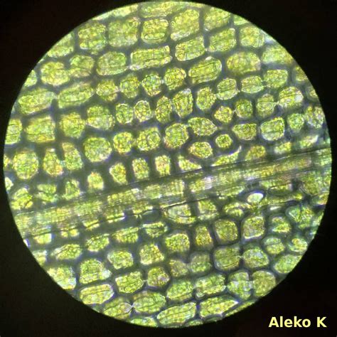
Table of Contents
What Three-Dimensional Shape Are Elodea Cells? Exploring the Microscopic World of Aquatic Plants
Elodea, a genus of aquatic plants commonly known as waterweeds, provides a captivating subject for microscopic observation. Their cells, easily visible under a light microscope, offer a fascinating glimpse into the fundamental building blocks of plant life. But what exactly is the three-dimensional shape of these cells? This question leads us on a journey into the intricacies of plant cell structure, the limitations of microscopy, and the interpretation of two-dimensional images in a three-dimensional context.
Understanding Elodea Cells: A Microscopic Perspective
Elodea cells are renowned for their large size and relatively simple structure, making them ideal for introductory microscopy studies. When viewed under a microscope, the most striking features are their rectangular or prismatic appearance. This rectangular shape, however, is only a two-dimensional projection of the cell's true three-dimensional structure. The apparent rectangularity is a result of the cell wall, a rigid outer layer providing structural support and shape to the cell.
The Cell Wall: The Defining Factor of Shape
The cell wall of Elodea cells is primarily composed of cellulose, a complex carbohydrate that forms a strong, yet flexible framework. This framework is responsible for the cell's relatively rigid shape, contributing significantly to the observed rectangular profile. However, the cell wall doesn't simply create a perfect rectangular prism; deviations from this idealized shape are common.
Cytoplasm and Organelles: Internal Structure and Influence on Shape
Inside the cell wall lies the cytoplasm, a gel-like substance containing various organelles, including chloroplasts, the sites of photosynthesis. The chloroplasts are clearly visible under a light microscope as small, oval, green structures that constantly stream around the cell's interior, a process known as cytoplasmic streaming or cyclosis. While the chloroplasts contribute to the cell's internal volume, their influence on the overall three-dimensional shape is less significant compared to the cell wall.
Vacuoles: Occupying the Cellular Space
Elodea cells also possess a large central vacuole, a membrane-bound sac filled with water and various dissolved substances. This vacuole plays a crucial role in maintaining cell turgor pressure—the pressure exerted by the cell contents against the cell wall. This turgor pressure is vital for maintaining the cell's shape and rigidity. The size of the vacuole can change based on water availability, subtly affecting the overall cell shape.
Moving Beyond Two Dimensions: Visualizing the 3D Structure
The challenge lies in translating the two-dimensional microscopic image into a comprehensive three-dimensional understanding. While a light microscope provides a clear view of the cell's surface, it offers only a limited perspective. To truly grasp the three-dimensional structure, we need to consider several points:
The Limitations of Microscopy
Light microscopy, though invaluable for observing Elodea cells, provides only a limited view of their structure. It presents a single plane of focus at any one time, making it difficult to simultaneously visualize the entire cell’s depth. We are essentially looking at a slice through the cell’s three-dimensional form.
Reconstructing the 3D Model
To obtain a more comprehensive 3D understanding, we would need to employ techniques beyond standard light microscopy. Advanced microscopy methods like confocal microscopy or 3D image reconstruction from serial sections (obtained using electron microscopy) would be required. These techniques allow for the visualization of the cell at multiple focal planes, enabling the construction of a three-dimensional model.
The Approximation: Prismatic Shape
Based on observations using light microscopy and the understanding of plant cell wall structure, we can conclude that the closest approximation of the three-dimensional shape of an Elodea cell is a slightly irregular prism. The irregularities arise from the inherent variability in cell wall formation and the influence of the internal cellular structures like the vacuole and streaming chloroplasts.
Factors Affecting Elodea Cell Shape: External Influences
While the inherent properties of the cell wall and internal structures primarily determine the Elodea cell's shape, external factors also play a role:
Water Availability and Turgor Pressure
As mentioned earlier, water availability directly affects the size of the central vacuole and the resulting turgor pressure. In a hypotonic environment (where the water potential outside the cell is higher than inside), water enters the cell, causing the vacuole to expand and the cell to become more turgid and its shape closer to a perfect prism. Conversely, in a hypertonic environment (where the water potential outside the cell is lower), water exits the cell, causing the vacuole to shrink, leading to a less rigid and possibly slightly shrunken cell shape.
Environmental Stressors
Environmental stress, such as extreme temperatures, nutrient deficiency, or exposure to toxins, can affect cell wall synthesis and integrity, leading to alterations in cell shape and size. Cells under stress may exhibit irregular shapes, deviating significantly from the idealized prismatic structure.
Age of the Cell
Young, actively growing Elodea cells may display slightly different shapes than mature cells. The cell wall undergoes continuous modification and remodeling during growth, influencing the final three-dimensional form. Older cells might exhibit some degree of cell wall thickening or even slight deformities.
Beyond the Rectangular Illusion: Deeper Understanding
The seemingly simple rectangular shape observed in Elodea cells under a light microscope masks a richer, more dynamic three-dimensional structure. Understanding this three-dimensional morphology requires going beyond the limitations of simple microscopy and acknowledging the influence of both internal cellular components and external environmental factors. By appreciating these complexities, we gain a deeper understanding of the intricate workings of plant cells and the factors that shape their form and function.
Exploring Further: Advanced Microscopy Techniques
While a basic light microscope suffices for initial observations, employing advanced techniques significantly enhances our understanding of Elodea cell shape. Confocal microscopy, for instance, creates high-resolution images by optically sectioning the specimen, allowing for the construction of detailed 3D models. This method provides invaluable insights into the cell's internal architecture, revealing the true three-dimensional arrangement of organelles within the cellular matrix.
Electron microscopy, while offering even higher resolution, requires sample preparation techniques that can potentially alter the cell's natural structure. However, electron microscopy, particularly transmission electron microscopy (TEM) allows for a detailed look at the ultrastructure of the cell wall, revealing the intricate network of cellulose microfibrils. This information helps explain the rigidity and structural characteristics of the cell wall, ultimately contributing to our understanding of the cell's shape.
Conclusion: The Dynamic Three-Dimensional Elodea Cell
In conclusion, the three-dimensional shape of an Elodea cell is best approximated as a slightly irregular prism. This shape is primarily determined by the rigid cell wall made of cellulose, but is subtly influenced by the internal structures—the central vacuole and the streaming chloroplasts—and significantly affected by environmental conditions and the cell's age. While a simple light microscope reveals a two-dimensional projection of this prism, advanced microscopy techniques offer a far more comprehensive view of its true three-dimensional complexity, revealing the dynamic interplay of factors influencing the form and function of these fascinating aquatic plant cells. Further research into the molecular mechanisms governing cell wall biosynthesis and the impact of various environmental stimuli on cell morphology would further enhance our understanding of this fundamental biological building block.
Latest Posts
Latest Posts
-
What Is Always True When There Is A Land Breeze
Mar 04, 2025
-
Sscg 15 A E Standard
Mar 04, 2025
-
Physicians Will Be Penalized By The Cms If They
Mar 04, 2025
-
Formal Article Or Book Based On Dental Evidence And Facts
Mar 04, 2025
-
Experiment 2 Qualitative Analysis Known And Unknown Ions
Mar 04, 2025
Related Post
Thank you for visiting our website which covers about What Three Dimensional Shape Are Elodea Cells . We hope the information provided has been useful to you. Feel free to contact us if you have any questions or need further assistance. See you next time and don't miss to bookmark.
