Activity 11 Optics Of The Human Eye
Onlines
Mar 03, 2025 · 7 min read
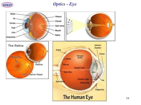
Table of Contents
Activity 11: Optics of the Human Eye: A Deep Dive into Vision
The human eye, a marvel of biological engineering, is a complex optical instrument responsible for our ability to perceive the world around us. Understanding its optical properties is crucial to appreciating the intricacies of vision and diagnosing vision problems. This comprehensive exploration of Activity 11, focusing on the optics of the human eye, will delve into the structures involved, the mechanisms of light refraction and image formation, common refractive errors, and the role of accommodation.
The Anatomy of Vision: Key Optical Components
The eye's optical system effectively works like a camera, capturing light and transforming it into a neural signal the brain interprets as an image. Several key structures play crucial roles in this process:
1. The Cornea: The Eye's First Lens
The cornea, a transparent, dome-shaped structure at the front of the eye, is the first refractive surface encountered by incoming light. Its curved surface bends (refracts) light rays, significantly contributing to the eye's focusing power. The cornea's refractive index is approximately 1.376, slightly higher than that of air, causing light rays to converge. Its fixed refractive power accounts for a substantial portion of the eye's total refractive power. Its clarity and smooth surface are essential for sharp vision; any irregularity can lead to blurred vision.
2. The Aqueous Humor: Filling the Anterior Chamber
Behind the cornea lies the anterior chamber, filled with aqueous humor, a watery fluid. This fluid maintains the shape of the cornea and provides nutrients to the lens and cornea. While the aqueous humor's refractive index (approximately 1.336) is relatively close to that of the cornea, it contributes to the overall refractive power of the eye. The constant production and drainage of aqueous humor are crucial for maintaining intraocular pressure. Disruptions to this balance can lead to glaucoma.
3. The Lens: Accommodation and Fine-Tuning
The lens, a transparent, biconvex structure suspended behind the iris by zonular fibers, is the eye's adjustable refractive component. Unlike the cornea, the lens's refractive power can be altered through a process called accommodation. The ciliary muscles surrounding the lens control its shape. When these muscles relax, the lens becomes flatter, reducing its refractive power; this is for focusing on distant objects. Conversely, when the ciliary muscles contract, the lens becomes more rounded, increasing its refractive power, enabling focus on nearby objects. This ability to adjust focus is crucial for clear vision at varying distances. The lens's refractive index varies slightly across its structure, contributing to its focusing ability. Presbyopia, the age-related loss of accommodation, is a common condition affecting the ability to focus on near objects.
4. The Vitreous Humor: Maintaining Shape and Clarity
The vitreous humor, a gel-like substance filling the space between the lens and the retina, makes up the majority of the eye's volume. It helps maintain the eye's spherical shape and keeps the retina in place. While its refractive index (approximately 1.336) is similar to that of the aqueous humor, it contributes to the overall optical properties of the eye. Its transparency is crucial for clear vision; opacities within the vitreous humor can lead to floaters or other vision disturbances.
5. The Retina: Light Detection and Image Formation
Finally, the retina is the light-sensitive layer lining the back of the eye. It contains millions of photoreceptor cells—rods (responsible for vision in low light) and cones (responsible for color vision and visual acuity)—that convert light into electrical signals. These signals are then transmitted to the brain via the optic nerve, creating the visual perception. The retina's position at the focal point of the eye's optical system is essential for sharp image formation. Any displacement of the retina can lead to blurred vision or other visual impairments.
Refraction and Image Formation: The Physics of Sight
The process of vision relies heavily on the principles of refraction. As light passes from one medium to another (e.g., from air to cornea), its speed changes, causing it to bend. The amount of bending depends on the refractive indices of the two media and the angle of incidence.
The eye's optical system refracts incoming light rays, bringing them to a focus on the retina. For a clearly focused image, the focal point must coincide with the retina. The combined refractive power of the cornea and lens determines the eye's overall refractive power, which is measured in diopters (D). A typical eye has a total refractive power of around 60D.
The process of image formation is inverted: the image formed on the retina is upside down and reversed. However, the brain processes this information and interprets the image correctly. The sharpness of the image depends on the accuracy of focus on the retina. Any deviation from this ideal focus leads to refractive errors.
Refractive Errors: Common Vision Problems
Refractive errors occur when the eye's refractive power does not precisely focus light on the retina. These errors can be corrected with corrective lenses (glasses or contact lenses) or refractive surgery.
1. Myopia (Nearsightedness):
In myopia, the eye is too long, or the refractive power is too strong, causing light to focus in front of the retina. This results in blurry distance vision. Myopia is corrected with concave lenses, which diverge light rays, effectively shifting the focal point back onto the retina.
2. Hyperopia (Farsightedness):
Hyperopia occurs when the eye is too short, or the refractive power is too weak, causing light to focus behind the retina. This results in blurry near vision, although distance vision might be relatively clear. Hyperopia is corrected with convex lenses, which converge light rays, bringing the focal point forward onto the retina.
3. Astigmatism:
Astigmatism is a condition where the cornea or lens has an irregular shape, leading to blurred vision at all distances. Different meridians of the eye have different refractive powers, causing multiple focal points instead of a single one. Astigmatism is corrected with cylindrical lenses that compensate for the irregular shape of the cornea or lens.
Accommodation: Adjusting Focus for Near and Far Vision
The lens's ability to change its shape, a process known as accommodation, is crucial for focusing on objects at different distances. As objects move closer, the ciliary muscles contract, making the lens more rounded and increasing its refractive power. This allows the eye to maintain a sharp focus on the near object. Conversely, as objects move farther away, the ciliary muscles relax, flattening the lens and reducing its refractive power. This adaptation is essential for clear vision at all distances. The amplitude of accommodation, the range over which the eye can effectively adjust its focus, decreases with age, leading to presbyopia.
Beyond the Basics: Advanced Concepts in Eye Optics
While this overview covers the fundamental aspects of the eye's optics, several more advanced concepts enhance our understanding:
-
Pupillary Light Reflex: The pupil's ability to constrict and dilate in response to changing light levels. This reflex regulates the amount of light entering the eye, protecting the retina from damage and optimizing image quality.
-
Chromatic Aberration: The eye's tendency to refract different wavelengths of light differently, leading to slight color fringes around objects. The eye partially compensates for this effect, but it can still contribute to slight image imperfections.
-
Diffraction: The spreading of light waves as they pass through the pupil. Diffraction limits the eye's resolving power, setting a fundamental limit on the smallest details that can be distinguished.
-
Optical Coherence Tomography (OCT): A sophisticated imaging technique that uses light waves to create detailed cross-sectional images of the retina and other eye structures. This non-invasive technology is widely used in ophthalmology for diagnosing and monitoring various eye conditions.
Conclusion: The Eye as a Sophisticated Optical System
The human eye is a remarkable optical instrument, showcasing the elegance and complexity of biological design. Its intricate structures, precise refractive properties, and adaptive mechanisms combine to create clear, detailed vision. Understanding the optics of the human eye is crucial for appreciating the marvels of vision and addressing vision problems effectively. From the basic principles of refraction and accommodation to the advanced concepts of pupillary reflexes and chromatic aberration, a comprehensive knowledge of the eye's optical system enhances our understanding of this remarkable sense. Continued research and technological advancements continually refine our knowledge of the eye and its function, leading to better diagnostic tools and treatments for various visual impairments.
Latest Posts
Latest Posts
-
Jane Ai Clinical Judgement Assessments Gray
Mar 03, 2025
-
To Kill A Mockingbird Chapter Summaries
Mar 03, 2025
-
Ap Cs A Unit 5 Progess Check
Mar 03, 2025
-
All The Answers On Drivesafeonline 6 Hours Georgia Quizzes Answers
Mar 03, 2025
-
Dod Initial Orientation And Awareness Training Answers
Mar 03, 2025
Related Post
Thank you for visiting our website which covers about Activity 11 Optics Of The Human Eye . We hope the information provided has been useful to you. Feel free to contact us if you have any questions or need further assistance. See you next time and don't miss to bookmark.
