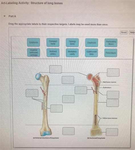
Art-labeling Activity Structure Of Long Bones
Onlines
Mar 31, 2025 · 7 min read

Table of Contents
Art-Labeling Activity Structure of Long Bones: A Comprehensive Guide
Art-labeling, or more formally, immunohistochemistry (IHC), is a powerful technique used to visualize the distribution of specific proteins within tissue samples. When applied to long bones, this technique provides invaluable insights into the complex processes of bone development, growth, remodeling, and repair. Understanding the intricacies of art-labeling activity within the different structural components of long bones is crucial for researchers in fields ranging from orthopedics and bone biology to pathology and regenerative medicine. This article delves into the detailed structural organization of long bones and how art-labeling helps us understand the dynamic processes occurring within each component.
The Structure of Long Bones: A Foundation for Understanding Art-Labeling
Long bones, such as the femur, tibia, and humerus, are characterized by their elongated shape and play crucial roles in locomotion and support. Their structure can be broadly categorized into several key components:
1. Diaphysis (Shaft):
The diaphysis constitutes the long, cylindrical shaft of the bone. It is primarily composed of compact bone, also known as cortical bone, which is dense and highly organized. Within the diaphysis, art-labeling studies often focus on:
-
Osteocytes: These bone cells reside within lacunae, interconnected by canaliculi. Art-labeling can reveal the expression of various proteins involved in bone metabolism, such as sclerostin, osteocalcin, and RANKL. Studying the distribution of these proteins can help understand bone remodeling and the response to mechanical loading.
-
Osteons (Haversian Systems): These cylindrical structures are the basic functional units of compact bone. They consist of concentric lamellae surrounding a central Haversian canal containing blood vessels and nerves. Art-labeling can identify the presence of specific proteins within osteons, providing insights into the processes of bone formation and resorption. For instance, identifying the presence of osteoprotegerin (OPG) could illuminate the bone's protective mechanisms against excessive bone resorption.
-
Interstitial Lamellae: These remnants of old osteons are located between intact osteons. Art-labeling can reveal the presence of proteins associated with bone remodeling, reflecting the dynamic nature of bone turnover.
2. Metaphysis:
The metaphysis is the region connecting the diaphysis to the epiphysis. It's a critical area of bone growth in children and adolescents. This region exhibits a distinct microstructure, characterized by:
-
Trabecular Bone (Cancellous Bone): Metaphyseal bone is primarily trabecular, a porous network of interconnected bony spicules. Art-labeling is particularly valuable in studying the metaphysis because the trabecular bone's high surface area facilitates rapid bone turnover. Researchers use art-labeling to visualize the distribution of growth factors (e.g., IGF-1, BMPs), signaling molecules (e.g., Wnt proteins), and other proteins involved in bone formation and growth plate function. Understanding the spatial distribution of these molecules can provide significant insights into the mechanisms of longitudinal bone growth.
-
Growth Plate (Epiphyseal Plate): This cartilaginous structure is responsible for the longitudinal growth of long bones. Art-labeling is crucial in investigating the cellular components of the growth plate (chondrocytes) and their maturation process. Studies can identify the expression of proteins associated with chondrocyte proliferation, differentiation, hypertrophy, and apoptosis, providing a detailed understanding of growth plate function and potential pathologies. For instance, visualizing the expression of type II collagen in the proliferative zone and type X collagen in the hypertrophic zone helps in understanding chondrocyte differentiation.
-
Vascularization: The metaphysis has a rich vascular supply, which plays a significant role in nutrient delivery and waste removal. Art-labeling can be used to study the distribution of vascular endothelial growth factor (VEGF) and other angiogenic factors, providing insights into the processes of bone vascularization and its relationship to bone growth and remodeling.
3. Epiphysis:
The epiphysis is the end portion of a long bone. In adults, it is primarily composed of:
-
Articular Cartilage: This specialized cartilage covers the articular surface, providing a smooth, low-friction surface for joint articulation. Art-labeling can be used to study the expression of cartilage-specific proteins like type II collagen and aggrecan, providing insights into cartilage health and the progression of osteoarthritis.
-
Subchondral Bone: This layer of bone lies beneath the articular cartilage. It plays a crucial role in transmitting forces across the joint. Art-labeling can reveal the presence of proteins associated with bone remodeling and the response to mechanical loading.
-
Trabecular Bone: Similar to the metaphysis, the epiphysis also contains trabecular bone, although the architecture differs. Art-labeling can help in studying the cellular activity and protein expression within this bone structure.
Art-Labeling Techniques in Long Bone Studies: Methods and Applications
Several art-labeling techniques are employed in long bone research, each with its unique advantages and limitations:
1. Immunofluorescence (IF):
IF uses fluorescently labeled antibodies to detect specific proteins. This technique allows for the visualization of multiple proteins simultaneously (multiplexing), providing a comprehensive view of the cellular interactions and molecular pathways involved in bone biology. Confocal microscopy is often used to obtain high-resolution images of labeled proteins within the complex three-dimensional structure of long bones. This technique is excellent for visualizing the location and distribution of proteins within specific cells and tissues.
2. Chromogenic Immunohistochemistry (IHC):
Chromogenic IHC employs enzyme-labeled antibodies, which produce a colored precipitate at the site of antigen binding. This method is widely used due to its relative simplicity and cost-effectiveness. While not as versatile as IF for multiplexing, it’s highly suitable for visualizing the localization of specific proteins in bone tissue sections.
3. In Situ Hybridization (ISH):
ISH is used to detect the presence of specific mRNA molecules within cells. This technique provides insights into the gene expression profiles of bone cells, complementing IHC data and providing a more comprehensive understanding of the molecular mechanisms regulating bone biology. By visualizing the mRNA for specific proteins, researchers can determine which genes are actively transcribed in different regions of the long bone.
Interpreting Art-Labeling Results: Challenges and Considerations
Interpreting art-labeling results requires careful consideration of several factors:
-
Antibody Specificity: The accuracy of the results depends heavily on the specificity of the antibodies used. Non-specific binding can lead to false-positive results.
-
Antigen Retrieval: The process of retrieving masked antigens is crucial for successful art-labeling. Different antigen retrieval methods may be necessary depending on the target protein and the fixation technique used.
-
Tissue Processing: The methods used to process and section the bone tissue can affect the preservation of antigens and the quality of the staining. Optimal tissue processing protocols are essential for reliable results.
-
Control Samples: Including positive and negative control samples is essential to validate the results and ensure the specificity of the art-labeling.
-
Quantification: Quantitative analysis of art-labeling results is often needed to determine the relative expression levels of proteins in different regions of the long bone. Image analysis software is often used to quantify the staining intensity and distribution.
Applications of Art-Labeling in Long Bone Research
Art-labeling plays a pivotal role in various aspects of long bone research:
1. Bone Development and Growth:
Art-labeling helps researchers understand the molecular mechanisms underlying bone growth, including the roles of growth factors, signaling pathways, and transcription factors. Studying the expression of various proteins in the growth plate allows for a deeper understanding of growth plate dysfunction and skeletal dysplasias.
2. Bone Remodeling and Repair:
Art-labeling helps to elucidate the cellular and molecular mechanisms involved in bone remodeling, including bone formation and resorption. Studying the spatial distribution of proteins involved in bone remodeling can help understand age-related bone loss and the development of osteoporosis.
3. Fracture Healing:
Art-labeling techniques are essential for studying the process of fracture healing, providing insights into the roles of different cell types and signaling molecules. This knowledge is crucial for developing novel strategies to enhance fracture healing.
4. Bone Diseases and Disorders:
Art-labeling is crucial in studying the pathogenesis of various bone diseases, including osteoporosis, osteosarcoma, and osteoarthritis. By identifying specific protein markers, researchers can improve diagnostic tools and therapeutic strategies.
Conclusion: The Future of Art-Labeling in Long Bone Research
Art-labeling techniques, such as IHC and IF, are indispensable tools for advancing our understanding of long bone biology. As technology advances, new and more sophisticated art-labeling techniques will continue to emerge, offering greater sensitivity, specificity, and resolution. Combined with other advanced imaging and analytical techniques, art-labeling will continue to play a pivotal role in unraveling the complexities of bone development, growth, remodeling, and repair, leading to breakthroughs in the diagnosis, treatment, and prevention of bone diseases and disorders. Further research leveraging advanced multiplexing techniques and integrating art-labeling with other ‘omics’ technologies (genomics, proteomics, metabolomics) will greatly expand our understanding of the intricate molecular networks governing bone biology and pathology. This will ultimately translate into more effective therapies and improved patient outcomes.
Latest Posts
Latest Posts
-
How To Know My Number On Glo
Apr 02, 2025
-
English Language And Composition Section 1 Answer Key
Apr 02, 2025
-
Summary Of Book 2 Of The Odyssey
Apr 02, 2025
-
Green Wave Company Plans To Own And Operate
Apr 02, 2025
-
Classify Each Histogram Using The Appropriate Descriptions
Apr 02, 2025
Related Post
Thank you for visiting our website which covers about Art-labeling Activity Structure Of Long Bones . We hope the information provided has been useful to you. Feel free to contact us if you have any questions or need further assistance. See you next time and don't miss to bookmark.
