Concept Map Major Muscles Of The Thigh
Onlines
Apr 07, 2025 · 6 min read
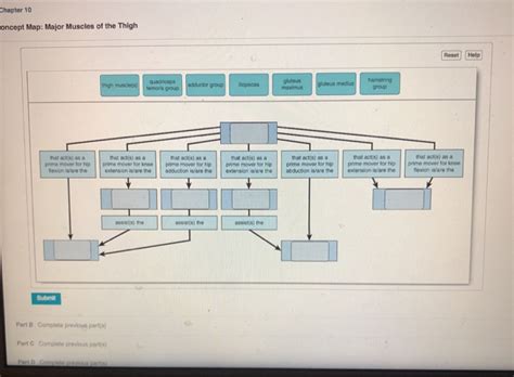
Table of Contents
Concept Map: Major Muscles of the Thigh
Understanding the complex network of muscles in the human thigh requires a structured approach. This article utilizes a concept map methodology to visually represent and explain the major thigh muscles, categorizing them by function and location. We will delve into the anatomy, actions, and clinical relevance of each muscle group, providing a comprehensive resource for students, athletes, and healthcare professionals alike.
I. Conceptual Overview: Dividing the Thigh Muscles
The thigh muscles can be broadly categorized into three compartments: the anterior (front), medial (inner), and posterior (back). This compartmentalization is crucial because muscles within the same compartment often share similar functions and innervations. Understanding this compartmentalization is the cornerstone of understanding thigh muscle function.
A. Anterior Compartment: Extensors of the Knee and Flexors of the Hip
This compartment primarily contains muscles responsible for extending the leg at the knee joint and flexing the thigh at the hip joint.
-
Quadriceps Femoris: This is the powerhouse of the anterior thigh, comprised of four distinct muscles:
-
Rectus Femoris: The only quadriceps muscle that crosses both the hip and knee joints. It flexes the hip and extends the knee. Origin: Anterior inferior iliac spine and superior acetabulum. Insertion: Tibial tuberosity via the patellar tendon. Clinical Relevance: Strains are common in athletes.
-
Vastus Lateralis: The largest of the quadriceps, located on the lateral side of the thigh. It extends the knee. Origin: Greater trochanter, intertrochanteric line, linea aspera. Insertion: Tibial tuberosity via the patellar tendon. Clinical Relevance: Intramuscular injections are frequently administered here.
-
Vastus Medialis: Situated on the medial side of the thigh. It extends the knee. Origin: Intertrochanteric line, linea aspera, medial supracondylar line. Insertion: Tibial tuberty via the patellar tendon. Clinical Relevance: Weakness can contribute to patellofemoral pain syndrome.
-
Vastus Intermedius: Deep to the rectus femoris, it extends the knee. Origin: Anterior and lateral surface of the femur. Insertion: Tibial tuberosity via the patellar tendon. Clinical Relevance: Often overlooked in clinical assessment.
-
-
Sartorius: The longest muscle in the body, it’s a weak flexor of the hip and knee, and laterally rotates the thigh. Origin: Anterior superior iliac spine. Insertion: Medial surface of the tibia. Clinical Relevance: Rarely injured individually but can be involved in more widespread injuries.
B. Medial Compartment: Adductors of the Thigh
The medial compartment muscles primarily adduct (bring the thigh towards the midline of the body).
-
Adductor Longus: Located superficially in the medial compartment. It adducts and flexes the thigh. Origin: Pubic symphysis. Insertion: Linea aspera of the femur. Clinical Relevance: Can be strained during athletic activities.
-
Adductor Brevis: Deep to the adductor longus, it adducts and flexes the thigh. Origin: Pubis. Insertion: Linea aspera of the femur. Clinical Relevance: Often injured in conjunction with other adductor muscles.
-
Adductor Magnus: The largest adductor muscle, with two heads (adductor and hamstring portions). It adducts, flexes, and extends the thigh (hamstring portion). Origin: Ischiopubic ramus, ischial tuberosity. Insertion: Linea aspera and adductor tubercle of the femur. Clinical Relevance: The most commonly injured adductor muscle.
-
Gracilis: A long, thin muscle that adducts the thigh and also assists in flexing the knee and medially rotating the leg. Origin: Pubic symphysis and inferior pubic ramus. Insertion: Medial surface of the tibia. Clinical Relevance: Frequently used in reconstructive surgery due to its length and strength.
C. Posterior Compartment: Extensors of the Hip and Flexors of the Knee (Hamstrings)
The posterior compartment contains the hamstring muscles, crucial for hip extension and knee flexion.
-
Biceps Femoris: Located on the lateral side of the thigh. It flexes the knee, extends the hip, and laterally rotates the leg. Origin: Ischial tuberosity (long head), linea aspera (short head). Insertion: Head of the fibula. Clinical Relevance: Prone to hamstring strains, particularly in athletes.
-
Semitendinosus: Located medially to the biceps femoris. It flexes the knee, extends the hip, and medially rotates the leg. Origin: Ischial tuberosity. Insertion: Medial surface of the tibia. Clinical Relevance: Often involved in hamstring injuries.
-
Semimembranosus: The deepest of the hamstrings, located medially to the semitendinosus. It flexes the knee, extends the hip, and medially rotates the leg. Origin: Ischial tuberosity. Insertion: Medial condyle of the tibia. Clinical Relevance: Similar injury patterns to semitendinosus.
II. Clinical Significance and Intermuscular Relationships
Understanding the functional relationships between these muscle groups is vital in clinical practice. Injuries often involve multiple muscles across compartments. For instance, a groin pull may involve the adductors and iliopsoas, while a hamstring strain might affect the biceps femoris, semitendinosus, and semimembranosus.
A. Common Injuries and their Muscular Basis
-
Hamstring Strains: These are frequent injuries, particularly in sprinting and jumping sports. They often involve a tear in one or more hamstring muscles, resulting in pain, swelling, and loss of function.
-
Quadriceps Strains: These strains often occur during sudden acceleration or deceleration, affecting the rectus femoris most commonly.
-
Adductor Strains: Adductor strains are common in sports requiring rapid changes in direction, such as soccer and hockey.
-
Patellofemoral Pain Syndrome: This condition involves pain around the kneecap and is often related to imbalances in the quadriceps and other muscles surrounding the knee.
B. Muscle Imbalances and their Implications
Imbalances between opposing muscle groups (e.g., quadriceps and hamstrings) can lead to various musculoskeletal problems. Weakness in one muscle group can overstress the opposing group, increasing the risk of injury. For example, weak hamstrings can increase the strain on the quadriceps during knee extension, increasing the risk of quadriceps strain.
C. Rehabilitation and Strengthening Strategies
Rehabilitation protocols for thigh muscle injuries often focus on restoring muscle strength, flexibility, and neuromuscular control. Specific exercises targeting each muscle group are crucial for effective rehabilitation. For example, strengthening exercises for the quadriceps might include leg extensions and squats, while hamstring exercises might include hamstring curls and glute bridges.
III. Advanced Considerations: Synergistic and Antagonistic Actions
The thigh muscles rarely act in isolation. Understanding their synergistic and antagonistic relationships provides a deeper insight into movement patterns.
A. Synergistic Muscle Actions
Synergistic muscles work together to produce a movement. For instance, during knee extension, the quadriceps muscles act synergistically. Similarly, several muscles contribute to hip flexion and adduction.
B. Antagonistic Muscle Actions
Antagonistic muscles work in opposition to each other. The quadriceps and hamstrings are a prime example of antagonistic muscle pairs; the quadriceps extend the knee while the hamstrings flex it. Proper balance between these antagonistic muscle groups is crucial for joint stability and injury prevention.
IV. Conclusion: A Holistic Approach to Thigh Muscle Anatomy
This concept map provides a structured overview of the major muscles of the thigh, categorized by compartment and function. Understanding the individual actions and interrelationships of these muscles is crucial for clinicians, athletes, and anyone interested in human movement. Furthermore, awareness of common injuries, muscle imbalances, and rehabilitation strategies allows for a more holistic and comprehensive approach to maintaining thigh muscle health and optimizing physical performance. This knowledge base empowers individuals to engage in targeted exercises and preventative measures to avoid injury and maintain optimal function. The integrated approach presented here emphasizes the interconnectedness of the various muscle groups and highlights the importance of a balanced and coordinated muscular system for efficient and injury-free movement.
Latest Posts
Latest Posts
-
Unit 8 Homework 6 Trigonometry Review
Apr 08, 2025
-
When A Mandated Reporter Finds A Family In Crisis
Apr 08, 2025
-
Discussion Thread How To Study The Genres Of The Bible
Apr 08, 2025
-
Disks Are Commonly Used With Sun Solaris Systems
Apr 08, 2025
-
A Lucky Individual Won The State Lottery
Apr 08, 2025
Related Post
Thank you for visiting our website which covers about Concept Map Major Muscles Of The Thigh . We hope the information provided has been useful to you. Feel free to contact us if you have any questions or need further assistance. See you next time and don't miss to bookmark.