Correctly Label The Following Anatomical Features Of The Thoracic Cavity
Onlines
Mar 22, 2025 · 6 min read
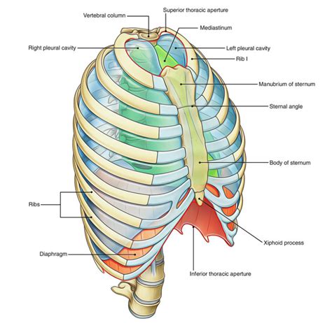
Table of Contents
Correctly Labeling the Anatomical Features of the Thoracic Cavity: A Comprehensive Guide
The thoracic cavity, also known as the chest cavity, is a vital part of the human body, housing crucial organs like the heart and lungs. Understanding its intricate anatomy is essential for anyone studying medicine, biology, or related fields. This comprehensive guide will delve into the key anatomical features of the thoracic cavity, providing detailed descriptions and guidance on correct labeling. We'll explore the bony structures, the membranes, the major organs, and their spatial relationships, ensuring a thorough understanding of this complex region.
I. The Bony Thorax: The Protective Cage
The thoracic cavity is primarily defined by the bony thorax, a strong, protective cage composed of several interconnected elements. Correctly labeling these components is fundamental to understanding the overall structure.
1. Sternum: The Breastbone
The sternum, or breastbone, is a flat, elongated bone located in the anterior midline of the thorax. It consists of three parts:
- Manubrium: The superior portion, articulating with the clavicles (collarbones) and the first two ribs.
- Body: The longest part, articulating with ribs 2-7.
- Xiphoid process: The small, inferior tip, often cartilaginous in younger individuals.
2. Ribs: The Bony Framework
Twelve pairs of ribs form the lateral walls of the thoracic cavity. They are classified into three groups based on their articulation with the sternum:
- True ribs (1-7): Directly connected to the sternum via their own costal cartilage.
- False ribs (8-10): Indirectly connected to the sternum, sharing costal cartilage with the rib above.
- Floating ribs (11-12): Do not connect to the sternum at all, ending freely in the abdominal musculature.
Each rib possesses a head, neck, tubercle, and angle, key anatomical landmarks used for identification and reference. Understanding the articulation points of each rib with the vertebrae is crucial for grasping the mechanics of respiration.
3. Thoracic Vertebrae: The Posterior Support
The thoracic vertebrae (T1-T12) form the posterior wall of the thoracic cavity. These vertebrae are unique in their size and shape, possessing features that distinguish them from cervical and lumbar vertebrae. Key features to identify include:
- Heart-shaped body: Larger than cervical vertebrae, and progressively larger towards the lumbar region.
- Long, slender spinous processes: Point inferiorly.
- Costal facets: Articulation points for the ribs, located on the vertebral bodies and transverse processes. These facets are essential for rib cage stability and movement.
II. Membranes of the Thoracic Cavity: A Protective Lining
The thoracic cavity is lined by a series of membranes that protect its internal organs and facilitate their movement. Accurate labeling of these membranes is crucial for understanding their functional roles.
1. Parietal Pleura: The Outer Lining
The parietal pleura is a serous membrane that lines the thoracic wall, the superior surface of the diaphragm, and the mediastinum. It's the outermost layer, providing a smooth, protective surface for the lungs and other structures.
2. Visceral Pleura: The Inner Lining
The visceral pleura directly adheres to the surface of each lung. It's continuous with the parietal pleura, forming a closed pleural sac around each lung. This sac creates a potential space, the pleural cavity, filled with a small amount of lubricating fluid. This fluid reduces friction during breathing.
3. Pericardium: The Heart's Protective Sac
The pericardium is a double-layered sac that surrounds the heart. It consists of two layers:
- Fibrous pericardium: The tough, outer layer that provides structural support.
- Serous pericardium: The inner layer, subdivided into parietal and visceral layers, with a pericardial cavity between them containing pericardial fluid. This fluid minimizes friction as the heart beats.
Understanding the relationship between the pericardium and the heart is critical in comprehending cardiac function and potential pathologies.
III. Major Organs of the Thoracic Cavity: The Heart and Lungs
The thoracic cavity houses two of the body’s most vital organs: the heart and lungs. Accurately labeling their components and their spatial relationships is paramount.
1. The Heart: The Body's Pump
The heart, a muscular organ roughly the size of a fist, is situated within the mediastinum, the central compartment of the thoracic cavity. Key features for labeling include:
- Apex: The inferior, pointed tip.
- Base: The superior, broader part.
- Atria (right and left): The receiving chambers.
- Ventricles (right and left): The pumping chambers.
- Major blood vessels: Aorta, vena cavae, pulmonary artery, and pulmonary veins.
2. The Lungs: The Respiratory Engines
The lungs are the primary organs of respiration, occupying the majority of the thoracic cavity on either side of the mediastinum. Key structures to identify include:
- Right lung: Typically larger and broader than the left lung, with three lobes.
- Left lung: Smaller and narrower than the right lung, with two lobes. The cardiac notch accommodates the heart's position.
- Bronchi: The branching airways leading into the lungs.
- Bronchioles: Smaller branches of the bronchi.
- Alveoli: Tiny air sacs where gas exchange occurs.
- Pulmonary vessels: Pulmonary arteries carry deoxygenated blood to the lungs, while pulmonary veins carry oxygenated blood back to the heart.
IV. Other Structures within the Thoracic Cavity
Beyond the heart and lungs, several other important structures reside within the thoracic cavity. These include:
- Trachea: The windpipe, carrying air to and from the lungs.
- Esophagus: The food pipe, transporting food from the pharynx to the stomach. It's located posterior to the trachea.
- Thymus gland: An endocrine gland, important in immune system development, especially during childhood.
- Great vessels: Major arteries and veins such as the aorta, vena cavae, and pulmonary vessels. These are crucial for blood circulation.
- Nerves: Numerous nerves pass through the thoracic cavity, innervating the organs and muscles of the region.
- Lymph nodes: Part of the lymphatic system, they filter lymph fluid and play a role in immunity.
V. Practical Application: Labeling Exercises and Resources
To solidify your understanding of the thoracic cavity's anatomy, consistent practice is key. Several approaches can enhance your learning:
- Anatomical models: Three-dimensional models offer a tangible way to visualize the relationships between structures. Careful observation and labeling of these models can significantly improve retention.
- Anatomical atlases: Detailed atlases provide high-quality images and descriptions. Using these in conjunction with labeling exercises will strengthen your understanding.
- Online resources: Many interactive online resources, such as virtual dissection tools, offer engaging and effective ways to learn anatomical structures.
VI. Clinical Significance: Understanding Thoracic Pathology
A thorough understanding of thoracic cavity anatomy is essential for diagnosing and treating various medical conditions. For example, knowledge of the pleural space is crucial for understanding pleural effusions (fluid build-up) or pneumothorax (collapsed lung). Similarly, an understanding of the cardiac anatomy is critical for diagnosing and managing heart conditions.
Thoracic pathologies can involve any of the structures discussed here, highlighting the interconnectivity and importance of each component.
Conclusion: Mastery through Practice and Application
Correctly labeling the anatomical features of the thoracic cavity requires dedication and focused learning. By combining the study of text-based resources with hands-on experiences using models and interactive tools, you can build a strong foundation in this vital area of anatomy. Remember, consistent practice, combined with a clear understanding of the functional relationships between structures, will lead to mastery. The detailed information provided in this guide serves as a stepping stone towards a comprehensive understanding of this complex and crucial region of the human body. Further exploration into specialized anatomical texts and clinical case studies will enhance your knowledge and ability to apply this information in a practical context.
Latest Posts
Latest Posts
-
Which Evasion Aids Can Assist You
Mar 24, 2025
-
Which Statement Related To Bonds Is True
Mar 24, 2025
-
Tina Jones Cardiovascular Shadow Health Subjective
Mar 24, 2025
-
Skills Module 3 0 Central Venous Access Devices Posttest
Mar 24, 2025
-
What Role Does Imc Play In Under Armor Marketing
Mar 24, 2025
Related Post
Thank you for visiting our website which covers about Correctly Label The Following Anatomical Features Of The Thoracic Cavity . We hope the information provided has been useful to you. Feel free to contact us if you have any questions or need further assistance. See you next time and don't miss to bookmark.
