Correctly Label The Following Anatomical Parts Of Osseous Tissue
Onlines
Mar 10, 2025 · 7 min read
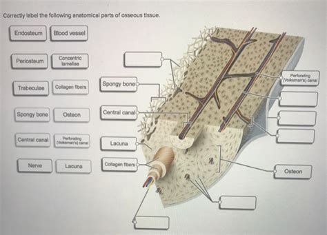
Table of Contents
Correctly Labeling the Anatomical Parts of Osseous Tissue
Understanding the anatomy of osseous tissue, commonly known as bone, is crucial for anyone studying biology, anatomy, or related fields. Bones are not simply inert structures; they are dynamic, living organs responsible for a multitude of functions, including support, protection, movement, mineral storage, and blood cell production. This article will provide a comprehensive guide to correctly labeling the various anatomical parts of osseous tissue, from the macroscopic level down to the microscopic components. We'll explore the different types of bone, their structures, and the cells that contribute to their formation, maintenance, and repair.
I. Macroscopic Anatomy of Bones: The Big Picture
Before diving into microscopic details, let's examine the overall structure of a typical long bone, which serves as a good model for understanding many key features found in other bone types.
A. Key External Features of a Long Bone:
-
Diaphysis: This is the long shaft of the bone. It's primarily composed of compact bone, providing strength and rigidity. The diaphysis is crucial for weight bearing and leverage during movement.
-
Epiphysis: Located at the ends of the diaphysis, these are the expanded portions of the bone. The epiphyses are primarily composed of spongy bone, which is lighter yet still strong enough to withstand forces. The articular surfaces of the epiphyses are covered with hyaline cartilage, facilitating smooth joint movement.
-
Metaphysis: This is the transitional region between the diaphysis and epiphysis. In growing bones, the metaphysis contains the epiphyseal plate (growth plate), a cartilaginous region responsible for longitudinal bone growth. Once growth is complete, the epiphyseal plate ossifies, leaving behind the epiphyseal line.
-
Periosteum: This is a tough, fibrous membrane covering the outer surface of the bone (except for the articular surfaces). It's richly supplied with blood vessels and nerves, providing nutrients and signaling for bone growth and repair. The periosteum also serves as an attachment point for tendons and ligaments.
-
Endosteum: This is a thin, connective tissue membrane lining the inner surface of the medullary cavity. It contains bone-forming cells (osteoblasts) and bone-resorbing cells (osteoclasts), playing a crucial role in bone remodeling.
-
Medullary Cavity: This is the hollow space within the diaphysis. In adults, it primarily contains yellow bone marrow, which is largely adipose tissue. In children, it contains red bone marrow, which is actively involved in hematopoiesis (blood cell formation).
B. Different Bone Types:
While long bones serve as a good example, bones come in various shapes and sizes, each adapted to its specific function:
-
Long Bones: Longer than they are wide (e.g., femur, humerus). Characterized by a diaphysis, epiphyses, and medullary cavity.
-
Short Bones: Approximately equal in length, width, and height (e.g., carpals, tarsals). Primarily composed of spongy bone.
-
Flat Bones: Thin and flattened, often curved (e.g., skull bones, ribs, scapula). Provide protection and broad surfaces for muscle attachment.
-
Irregular Bones: Complex shapes that don't fit into other categories (e.g., vertebrae, facial bones). Their structure reflects their diverse functions.
-
Sesamoid Bones: Small, round bones embedded within tendons (e.g., patella). They reduce friction and improve mechanical advantage.
II. Microscopic Anatomy of Bones: A Cellular Perspective
Now let's delve into the microscopic structure of bone tissue, focusing on the cells and matrix that make up this vital organ.
A. Components of the Bone Matrix:
-
Organic Components: These include collagen fibers and ground substance, providing flexibility and tensile strength to the bone. Collagen fibers are critical for resisting tension and preventing fractures. The ground substance contains various proteins and glycosaminoglycans that contribute to the bone's overall structure.
-
Inorganic Components: Primarily composed of calcium phosphate crystals (hydroxyapatite), these provide hardness and compressive strength to the bone. The interplay of organic and inorganic components ensures the bone's exceptional strength and resilience.
B. Bone Cells: The Architects and Maintainers
Several specialized cell types contribute to the formation, maintenance, and remodeling of bone tissue:
-
Osteoblasts: These are bone-forming cells. They synthesize and secrete the organic components of the bone matrix (osteoid), which then mineralizes to form new bone tissue. Osteoblasts are responsible for the initial formation of bone and its continued growth and repair.
-
Osteocytes: These are mature bone cells that reside within lacunae (small spaces) within the bone matrix. They are derived from osteoblasts that have become embedded in the matrix. Osteocytes play a crucial role in maintaining bone tissue, sensing mechanical stress, and regulating bone remodeling. They communicate with each other and with osteoblasts and osteoclasts via canaliculi (small canals).
-
Osteoclasts: These are large, multinucleated cells responsible for bone resorption (breakdown of bone tissue). They secrete acids and enzymes that dissolve the mineral and organic components of the bone matrix. This process is essential for bone remodeling, calcium homeostasis, and repair of damaged bone.
-
Bone Lining Cells: These are quiescent (inactive) cells that cover the surfaces of bone that are not undergoing remodeling. They help to maintain the integrity of the bone surface and may play a role in regulating bone remodeling.
C. Microscopic Structures:
-
Compact Bone: This type of bone is dense and solid, forming the outer layer of most bones and the bulk of the diaphysis of long bones. It's organized into osteons (Haversian systems), which are cylindrical units containing concentric lamellae (rings) of bone matrix surrounding a central Haversian canal. The Haversian canals contain blood vessels and nerves, providing nutrients and communication to the osteocytes within the osteons. Volkmann's canals are perpendicular to the Haversian canals, connecting them to the periosteum and endosteum.
-
Spongy Bone (Cancellous Bone): This type of bone is lighter and less dense than compact bone, consisting of a network of trabeculae (thin, bony plates) and interconnected spaces filled with bone marrow. Spongy bone is found in the epiphyses of long bones and in the interior of many other bones. Its porous structure allows for lightweight yet strong support, and the spaces within contribute to blood cell formation in red bone marrow.
III. Clinical Significance: Understanding Bone Disorders
Correctly labeling the anatomical parts of osseous tissue is not only important for academic understanding but also has significant clinical implications. Many diseases and disorders affect the bones, and accurate diagnosis and treatment often rely on a thorough understanding of bone anatomy.
A. Fractures:
Fractures, or breaks in the bone, can occur in various parts of the bone and are classified according to their location and type (e.g., transverse, oblique, spiral, comminuted, greenstick). Proper identification of the fracture site is crucial for effective treatment and recovery.
B. Osteoporosis:
This condition is characterized by decreased bone mass and density, making bones more susceptible to fractures. Understanding bone microstructure, including the balance between osteoblast and osteoclast activity, is essential for diagnosing and managing osteoporosis.
C. Osteoarthritis:
This degenerative joint disease affects the articular cartilage covering the epiphyses of bones, leading to pain, stiffness, and reduced mobility. Understanding the structure and function of the articular cartilage and its relationship to underlying bone is crucial for diagnosis and treatment.
D. Bone Tumors:
Both benign and malignant tumors can develop in bones, affecting various parts of the bone structure. Accurate labeling and identification of the location of the tumor are essential for appropriate treatment planning.
IV. Conclusion: The Importance of Accurate Labeling
Correctly labeling the anatomical parts of osseous tissue is fundamental to understanding the structure, function, and clinical significance of bone. From the macroscopic features of long bones to the microscopic details of bone cells and matrix, each component plays a vital role in maintaining bone health and overall body function. A comprehensive knowledge of this complex tissue is essential for healthcare professionals, researchers, and anyone interested in the fascinating world of human anatomy and physiology. The detailed understanding of bone structure, down to the cellular level, allows for a better understanding of its intricate processes and facilitates advancements in diagnosis and treatment of bone-related pathologies. Continuous research expands our knowledge, leading to more effective methods for preventing and treating bone diseases, improving quality of life for individuals affected by these conditions.
Latest Posts
Latest Posts
-
Your Patient Is Experiencing Left Sided Weakness From A Meningioma
Mar 11, 2025
-
A 12 Year Old Male Jumped Approximately
Mar 11, 2025
-
Formulas And Nomenclature Binary Ionic Compounds Worksheet
Mar 11, 2025
-
1 3 Skills Practice Locating Points And Midpoints Answer Key
Mar 11, 2025
-
Service Based Gifts Are Important Because
Mar 11, 2025
Related Post
Thank you for visiting our website which covers about Correctly Label The Following Anatomical Parts Of Osseous Tissue . We hope the information provided has been useful to you. Feel free to contact us if you have any questions or need further assistance. See you next time and don't miss to bookmark.
