Correctly Label The Following External Anatomy Of The Anterior Heart
Onlines
Mar 26, 2025 · 6 min read
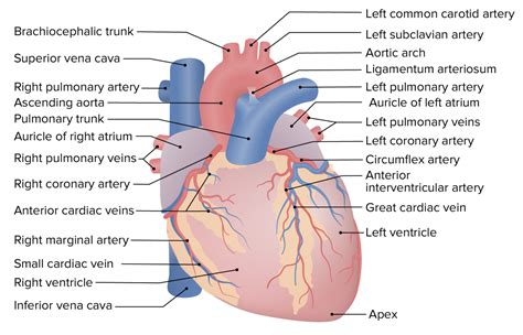
Table of Contents
Correctly Label the Following External Anatomy of the Anterior Heart
Understanding the external anatomy of the anterior heart is fundamental to comprehending cardiovascular function. This detailed guide will walk you through correctly labeling the key structures visible on the anterior surface of the heart, providing comprehensive descriptions and clinical significance for each. Mastering this knowledge is crucial for medical students, healthcare professionals, and anyone interested in the intricacies of human anatomy and physiology.
Key Structures of the Anterior Heart: A Detailed Guide
The anterior surface of the heart presents several distinct features. Correctly identifying these structures requires careful observation and a strong understanding of their relative positions and functions. Let's delve into each structure individually:
1. Right Atrium (RA)
-
Location: The right atrium is located on the right side of the heart, forming a significant portion of the anterior surface. It's the receiving chamber for deoxygenated blood returning from the body through the superior and inferior vena cava.
-
Identification: The RA is typically identified by its relatively smooth surface compared to the rougher texture of the right ventricle. You might observe the superior and inferior vena cava entering the atrium.
-
Clinical Significance: Conditions affecting the right atrium, such as atrial fibrillation or enlargement (right atrial dilatation), can have serious implications for blood flow and overall cardiovascular health.
2. Right Ventricle (RV)
-
Location: The right ventricle is positioned inferior and slightly anterior to the right atrium. It forms a substantial portion of the anterior cardiac surface.
-
Identification: The RV is characterized by its thicker muscular wall compared to the RA. You might observe the pulmonary trunk emerging from its superior aspect. The presence of trabeculae carneae (muscular ridges) on the internal surface is a distinctive feature.
-
Clinical Significance: The RV plays a critical role in pumping deoxygenated blood to the lungs. Conditions like pulmonary hypertension can significantly stress the RV, leading to right ventricular failure.
3. Left Ventricle (LV)
-
Location: Although mostly posterior, a portion of the left ventricle contributes to the anterior heart surface, particularly at the apex.
-
Identification: The LV is generally less visible anteriorly than the RV and RA. It's identified by its much thicker muscular wall than the right ventricle. This is because it needs to generate significantly higher pressure to pump oxygenated blood to the systemic circulation.
-
Clinical Significance: The LV is the powerhouse of the heart. Its efficiency is critical for maintaining adequate systemic blood flow. Conditions like left ventricular hypertrophy or left ventricular failure have profound implications for overall health.
4. Pulmonary Trunk (PT)
-
Location: The pulmonary trunk is a large artery originating from the right ventricle, positioned superiorly and slightly anteriorly on the heart's anterior surface.
-
Identification: The PT is readily identifiable by its relatively large diameter and its position immediately superior to the right ventricle. It branches into the left and right pulmonary arteries.
-
Clinical Significance: Obstruction of the pulmonary trunk, such as by a pulmonary embolism, can dramatically impair blood flow to the lungs and cause respiratory distress.
5. Pulmonary Artery (PA)
-
Location: Immediately branching from the pulmonary trunk are the left and right pulmonary arteries. The main pulmonary artery's bifurcation is visible on the anterior surface.
-
Identification: These arteries are easier to identify once the pulmonary trunk is located. They are identified as the vessels carrying deoxygenated blood away from the heart towards the lungs for oxygenation.
-
Clinical Significance: Conditions affecting the pulmonary arteries, like pulmonary embolism or pulmonary hypertension, can lead to significant respiratory and cardiovascular problems.
6. Superior Vena Cava (SVC)
-
Location: The SVC is a large vein returning deoxygenated blood from the upper body to the right atrium. It enters the heart on the superior aspect, contributing to the anterior view.
-
Identification: The SVC is large and easily distinguished from the other vessels. Its entrance into the right atrium is often clearly visible.
-
Clinical Significance: Obstruction of the SVC can lead to significant swelling in the upper body due to impaired venous return.
7. Inferior Vena Cava (IVC)
-
Location: The IVC is a large vein returning deoxygenated blood from the lower body to the right atrium. Although mainly posterior, a portion can be seen on the anterior aspect, especially when the heart is viewed at a slightly oblique angle.
-
Identification: Similar to the SVC, the IVC is large and readily identifiable by its location and direction of blood flow.
-
Clinical Significance: Conditions affecting the IVC, such as thrombosis, can lead to significant lower extremity swelling and potential pulmonary embolism.
8. Aorta (Ascending Aorta)
-
Location: The ascending aorta, the initial portion of the aorta, originates from the left ventricle. A small part of its origin may be visible from an anterior view.
-
Identification: The aorta is a large, thick-walled artery. While mostly posterior, a portion of its base is often visible near the pulmonary trunk.
-
Clinical Significance: Aortic diseases, including aneurysms or stenosis, are extremely serious and can be life-threatening.
9. Coronary Sulcus (Atrioventricular Groove)
-
Location: This groove encircles the heart, separating the atria from the ventricles. A significant portion of the coronary sulcus is visible on the anterior heart surface.
-
Identification: The coronary sulcus appears as a clear depression across the heart's surface. The major coronary arteries lie within this groove.
-
Clinical Significance: The location of the coronary arteries within the coronary sulcus makes this area critically important in understanding coronary artery disease.
10. Anterior Interventricular Sulcus
-
Location: This sulcus runs along the anterior surface of the heart, separating the right and left ventricles.
-
Identification: This is a visible groove running down the front of the heart. The anterior interventricular artery lies within this sulcus.
-
Clinical Significance: The anterior interventricular artery supplies a significant portion of the ventricles; therefore, blockages in this artery can cause significant damage to the heart muscle.
Clinical Correlations and Applications
Accurate identification of these structures is paramount in several clinical settings. For instance:
- Echocardiography: Echocardiograms rely heavily on the ability to visualize and measure these structures to assess cardiac function and diagnose various heart conditions.
- Cardiac Catheterization: This invasive procedure requires precise knowledge of the heart's anatomy to navigate catheters through the chambers and vessels.
- Cardiac Surgery: Surgeons must possess a deep understanding of the heart's external anatomy for successful cardiac interventions.
- Emergency Medicine: Rapid assessment of the heart's external anatomy is essential in trauma situations or during cardiac arrest.
Beyond the Anterior Surface: A Holistic Approach
While this guide focuses on the anterior heart, understanding the complete three-dimensional anatomy of the heart is crucial. The posterior surface contains additional vital structures like the left atrium, pulmonary veins, and the posterior descending artery. A thorough understanding of the entire heart's anatomy requires studying all surfaces.
Advanced Learning and Resources
To further enhance your understanding of the anterior heart's anatomy, consider exploring advanced resources such as:
- Anatomy textbooks: Detailed anatomical atlases provide comprehensive descriptions and high-quality images of the heart.
- Interactive anatomy software: Programs with 3D models allow for interactive exploration and manipulation of the heart's structures.
- Online anatomy resources: Many websites offer high-resolution images, animations, and interactive quizzes to aid in learning.
Mastering the external anatomy of the anterior heart is a journey of continuous learning. Regular revision, active recall, and practical application through anatomical models and clinical correlations will solidify your knowledge and enhance your ability to understand the complex workings of the human cardiovascular system. By combining meticulous study with clinical context, you will not only correctly label these structures but also understand their crucial role in maintaining human life. Remember, consistent effort and engagement with the subject matter are key to achieving a comprehensive understanding.
Latest Posts
Latest Posts
-
Characters In Do Androids Dream Of Electric Sheep
Mar 29, 2025
-
A Small Compact Car Was Involved In A Rollover Crash
Mar 29, 2025
-
Sick Call At Detention Facilities Must Be Held
Mar 29, 2025
-
Lewis Med Surg 12th Edition Test Bank
Mar 29, 2025
-
While Transferring A Patient To Als Staff Interference Should Be
Mar 29, 2025
Related Post
Thank you for visiting our website which covers about Correctly Label The Following External Anatomy Of The Anterior Heart . We hope the information provided has been useful to you. Feel free to contact us if you have any questions or need further assistance. See you next time and don't miss to bookmark.
