Correctly Label The Following Muscles Of The Anterior View
Onlines
Mar 12, 2025 · 7 min read
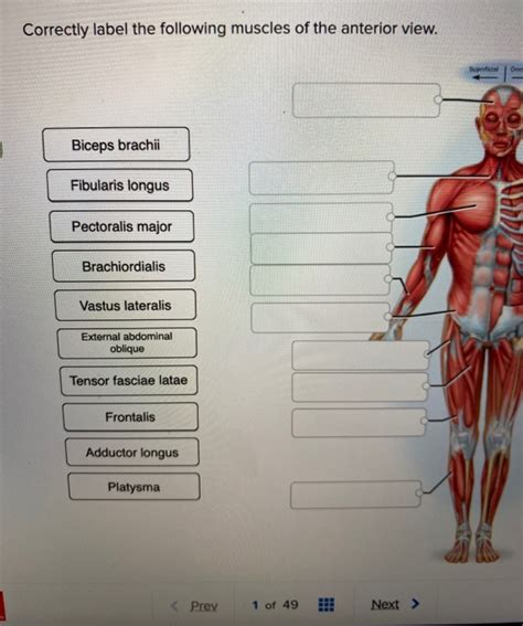
Table of Contents
Correctly Labeling the Muscles of the Anterior View: A Comprehensive Guide
Understanding the muscles of the human body, particularly from the anterior (front) view, is crucial for anyone studying anatomy, physiology, kinesiology, or related fields. This detailed guide will walk you through the correct labeling of the major anterior muscles, providing not only their names but also their functions and key characteristics. We'll cover everything from superficial muscles easily visible to deeper layers requiring a more in-depth understanding. This comprehensive approach will help you build a strong foundation in human anatomy and improve your understanding of movement and function.
The Superficial Muscles of the Anterior View
Let's start with the muscles most readily visible when observing the anterior view of the human body. These superficial muscles often play a significant role in large movements and are easier to identify due to their size and location.
1. Pectoralis Major:
- Location: This large, fan-shaped muscle covers a significant portion of the chest. It originates from the clavicle, sternum, and ribs and inserts on the humerus (upper arm bone).
- Function: The pectoralis major is primarily responsible for adduction (bringing the arm towards the body), flexion (raising the arm forward), and medial rotation of the humerus. It also plays a role in forced inspiration (breathing in).
- Key Identifying Features: Its substantial size and its fan-like shape spreading from the sternum to the humerus make it easily recognizable.
2. Deltoid:
- Location: Situated on the shoulder, the deltoid is a thick, triangular muscle covering the shoulder joint.
- Function: The deltoid is a powerful abductor (raising the arm away from the body) and plays a crucial role in shoulder flexion, extension, and rotation. It's divided into three parts: anterior (front), medial (middle), and posterior (rear), each contributing to different movements.
- Key Identifying Features: Its prominent, rounded shape makes it stand out on the shoulder. You can easily palpate (feel) its fibers.
3. Biceps Brachii:
- Location: Located on the anterior aspect of the upper arm, the biceps brachii is a two-headed muscle (hence the name "biceps").
- Function: The biceps brachii is the primary flexor (bends the elbow) of the forearm and also assists in supination (turning the palm upwards).
- Key Identifying Features: Its two distinct heads (long and short) are easily visible and palpable, especially when flexing the elbow. The prominent biceps tendon is also a key feature.
4. Brachialis:
- Location: Deep to the biceps brachii, the brachialis is a powerful flexor of the elbow.
- Function: Although less visible than the biceps, the brachialis is a crucial elbow flexor, providing most of the force for this movement.
- Key Identifying Features: It's located immediately beneath the biceps brachii, and its bulk can be appreciated by deep palpation.
5. Brachioradialis:
- Location: Located on the lateral (outside) aspect of the forearm, the brachioradialis runs from the humerus to the radius (forearm bone).
- Function: It acts as a flexor of the elbow, particularly when the forearm is in a neutral position (neither supinated nor pronated).
- Key Identifying Features: Its location on the lateral side of the forearm and its relatively superficial position make it easier to identify than some deeper muscles.
6. Rectus Abdominis:
- Location: The "six-pack" muscle, the rectus abdominis runs vertically down the anterior abdomen.
- Function: It flexes the trunk (bending forward), assists in lateral flexion (bending sideways), and helps to stabilize the spine.
- Key Identifying Features: The characteristic "six-pack" appearance due to tendinous intersections is a distinctive feature.
7. External Oblique:
- Location: Situated laterally on the abdomen, the external oblique is the outermost of the abdominal muscles.
- Function: It assists in trunk flexion, lateral flexion, and rotation. It also plays a role in forced expiration (breathing out).
- Key Identifying Features: Its fibers run inferomedially (downward and towards the midline) creating a characteristic diagonal pattern.
8. Internal Oblique:
- Location: Deep to the external oblique, the internal oblique has fibers running perpendicular to the external oblique.
- Function: Similar to the external oblique, it assists in trunk flexion, lateral flexion, and rotation.
- Key Identifying Features: Its fibers are less visible than the external oblique, and its location deep to the external oblique makes it harder to visualize on a superficial view.
9. Transversus Abdominis:
- Location: The deepest of the abdominal muscles, the transversus abdominis runs horizontally across the abdomen.
- Function: It compresses the abdominal contents and plays a vital role in stabilizing the spine.
- Key Identifying Features: It is generally not visible on a superficial view, requiring a deeper anatomical understanding to identify.
10. Iliopsoas:
- Location: This muscle group, composed of the iliacus and psoas major, is a deep hip flexor. While portions may be visible depending on body fat percentage and abdominal musculature, it’s largely deep.
- Function: It powerfully flexes the hip joint (bringing the thigh towards the abdomen).
- Key Identifying Features: Its location deep within the pelvis makes it difficult to observe in a standard anterior view.
11. Sartorius:
- Location: The longest muscle in the body, the sartorius runs diagonally across the thigh.
- Function: It flexes, abducts, and laterally rotates the hip, and it also assists in knee flexion.
- Key Identifying Features: Its long, strap-like appearance and its diagonal course across the anterior thigh makes it easily recognizable.
12. Quadriceps Femoris:
- Location: This group of four muscles (rectus femoris, vastus lateralis, vastus medialis, and vastus intermedius) covers the anterior thigh.
- Function: The primary function is knee extension (straightening the leg). The rectus femoris also flexes the hip.
- Key Identifying Features: The powerful bulk of these muscles is readily apparent on the anterior thigh. The rectus femoris can be distinguished by its origin on the pelvis.
13. Tibialis Anterior:
- Location: Situated on the anterior aspect of the lower leg, the tibialis anterior is a key muscle of the lower leg.
- Function: It dorsiflexes (lifts the foot upwards) and inverts (turns the sole of the foot inwards) the foot.
- Key Identifying Features: Its location on the anterior shin makes it readily visible and palpable.
Deeper Muscles of the Anterior View
Moving beyond the superficial layers, we encounter deeper muscles that often work in coordination with the superficial muscles to produce complex movements. These muscles are harder to visualize without dissection or advanced imaging techniques.
14. Pectoralis Minor:
- Location: Located deep to the pectoralis major, the pectoralis minor is a smaller muscle that originates from the ribs and inserts on the scapula (shoulder blade).
- Function: It protracts (moves forward) and depresses the scapula.
- Key Identifying Features: It's difficult to visualize on a surface view and requires a more in-depth understanding of the underlying anatomy.
15. Serratus Anterior:
- Location: Located on the lateral chest wall, the serratus anterior originates from the ribs and inserts on the scapula.
- Function: It protracts and upwardly rotates the scapula, playing an important role in shoulder movements. It also helps stabilize the scapula against the rib cage.
- Key Identifying Features: The muscle is often visible as a series of digitations (tooth-like projections) along the lateral chest wall, particularly in individuals with low body fat.
Importance of Correct Muscle Labeling
Accurately labeling the muscles of the anterior view is crucial for several reasons:
- Understanding Movement: Knowing the origin, insertion, and function of each muscle allows you to understand how movements are generated.
- Clinical Applications: For healthcare professionals, accurate muscle identification is essential for diagnosis and treatment of injuries and other conditions.
- Exercise and Fitness: Understanding muscle anatomy helps in designing effective exercise programs that target specific muscles for improved strength and performance.
- Artistic Representation: Accurate anatomical knowledge is vital for artists aiming to portray the human body realistically.
Practical Tips for Learning Muscle Anatomy
Learning muscle anatomy requires dedication and practice. Here are some helpful tips:
- Visual Aids: Use anatomical charts, atlases, and models to visualize the muscles and their relationships.
- Palpation: Practice palpating (feeling) the muscles on yourself and others to get a better sense of their location and shape.
- Active Learning: Engage in activities that require movement, paying attention to the muscles involved.
- Mnemonics: Create memory aids (mnemonics) to remember the names and functions of the muscles.
- Progressive Learning: Start with the superficial muscles and gradually move to the deeper layers as your knowledge expands.
By consistently applying these strategies and using the detailed information provided in this guide, you will be well on your way to mastering the intricate anatomy of the anterior view. Remember, consistent study and practical application are key to retaining and understanding this complex subject. Further research into specific muscles and their interactions with other muscles within the body will only enhance your comprehension and knowledge.
Latest Posts
Latest Posts
-
Introduction To Systems Thinking D372
Mar 13, 2025
-
Where Does A Squirrel Keep Its Winter Clothes
Mar 13, 2025
-
Apex Nih Stroke Scale Test Group A
Mar 13, 2025
-
China Dolls Book Provided By Consumers In Sociology
Mar 13, 2025
-
Chapter 16 1 Measuring And Recording Vital Signs
Mar 13, 2025
Related Post
Thank you for visiting our website which covers about Correctly Label The Following Muscles Of The Anterior View . We hope the information provided has been useful to you. Feel free to contact us if you have any questions or need further assistance. See you next time and don't miss to bookmark.
