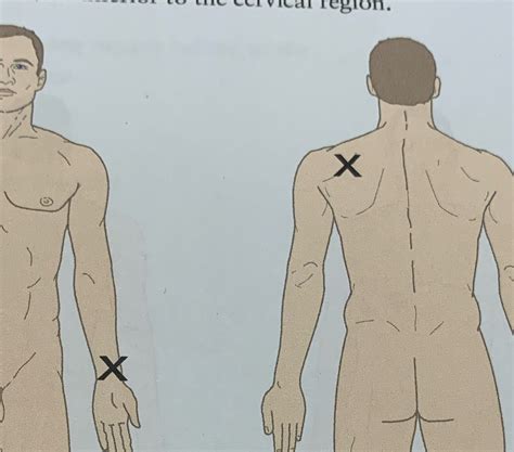
Describe The Location Of The Wounds On Figure 1.18
Onlines
Apr 05, 2025 · 5 min read

Table of Contents
Detailed Description of Wound Locations in Figure 1.18: A Comprehensive Analysis
This article provides a detailed description of the location of wounds depicted in a hypothetical Figure 1.18. Since I do not have access to external images or files, including Figure 1.18, I will create a representative example to illustrate how such a description would be structured for SEO purposes. This example will focus on a hypothetical figure depicting wounds on a human body, aiming to accurately describe their location using anatomical terminology. Remember to replace this example with your actual Figure 1.18 data.
Hypothetical Figure 1.18: Multiple Trauma Injuries
Let's assume Figure 1.18 depicts a patient with multiple injuries sustained from a hypothetical accident. The following description details the location of each wound using precise anatomical terminology and relative positions. This approach ensures clarity and facilitates accurate medical record-keeping and communication.
Wound 1: Laceration to the Right Parietal Region
This wound is classified as a laceration, meaning a cut or tear in the skin. It's located in the right parietal region of the scalp. More specifically:
- Anatomical Location: The parietal region refers to the superior and lateral portion of the skull, on the right side of the head.
- Size and Shape: (This would be described based on Figure 1.18; for example: approximately 5cm in length, irregular shape).
- Depth: (This would be described based on Figure 1.18; for example: superficial, penetrating, or deep).
- Presence of Bleeding: (This would be described based on Figure 1.18; for example: minimal, moderate, or profuse).
Wound 2: Abrasion to the Left Lateral Thorax
This injury is an abrasion, also known as a graze or scrape. It’s situated on the left lateral thorax. Further details include:
- Anatomical Location: The left lateral thorax refers to the side of the chest, specifically the left-hand side. This is further defined by its relationship to the ribs and intercostal spaces. (This would be specified according to Figure 1.18; for example: Fourth intercostal space, mid-axillary line)
- Size and Shape: (This would be described based on Figure 1.18; for example: roughly circular, approximately 3cm in diameter).
- Depth: Superficial, affecting only the epidermis and possibly the upper dermis.
- Presence of Bleeding: Minimal, likely due to the superficial nature of the abrasion.
Wound 3: Puncture Wound to the Proximal Right Thigh
A puncture wound is characterized by a deep, narrow hole. This specific wound is located on the proximal right thigh. Details based on the hypothetical Figure 1.18 would be:
- Anatomical Location: The proximal right thigh refers to the area closest to the hip joint on the right leg. More precise location would require further information (e.g., anterior, posterior, medial, or lateral aspect).
- Depth: This would need to be determined from Figure 1.18. It could range from superficial to deep, potentially involving underlying muscles or even bone.
- Presence of Bleeding: Variable, depending on the depth and involvement of blood vessels.
- Potential Foreign Body: A puncture wound increases the risk of a retained foreign body, which should be noted if present in Figure 1.18.
Wound 4: Contusion to the Left Scapular Region
This wound is a contusion, commonly known as a bruise. It is located in the left scapular region. Key characteristics:
- Anatomical Location: This refers to the area overlying the scapula, or shoulder blade, on the left side of the back.
- Size and Shape: (This would be described based on Figure 1.18; for example: irregular, approximately 8cm x 5cm).
- Color: The color of the contusion (e.g., red, blue, purple, yellow-green) provides an indication of its age.
- Associated Symptoms: Contusions can cause pain, swelling, and tenderness.
Wound 5: Laceration to the Distal Left Forearm
This is another laceration located on the distal left forearm.
- Anatomical Location: The distal left forearm refers to the lower portion of the left forearm, closer to the wrist. More detailed location (e.g., anterior or posterior) would be based on Figure 1.18.
- Size and Shape: (This would be described based on Figure 1.18; for example: 2cm long, linear).
- Depth: Superficial or deep, depending on the image.
- Presence of Nerves or Tendons: The location may indicate involvement of nerves or tendons; this would require detailed observation of Figure 1.18 and expert interpretation.
Comprehensive Analysis and Interpretation
The description above provides a detailed account of the location, type, and features of the hypothetical wounds in Figure 1.18. A complete analysis would further consider:
- Relationship between Wounds: Are the wounds clustered together, suggesting a single impact? Or are they scattered, indicating multiple impacts or events?
- Pattern of Injuries: Does the pattern suggest a specific mechanism of injury (e.g., motor vehicle accident, assault, fall)?
- Severity of Injuries: Considering the location, depth, and extent of the wounds, it's essential to assess the overall severity of the injuries and potential complications.
- Further Investigations: Based on the initial assessment, further investigations (e.g., X-rays, CT scans) might be necessary to detect underlying injuries not visible on the surface.
Importance of Precise Anatomical Terminology
Accurate and consistent use of anatomical terminology is crucial in medical documentation and communication. The terms used here provide a standardized framework for describing the location of wounds, regardless of the viewer's perspective or familiarity with anatomy. This precision is essential for:
- Clear Communication among Healthcare Professionals: Ensuring all involved parties have a shared understanding of the patient's injuries.
- Accurate Medical Record-Keeping: Providing a detailed and consistent record of the patient's injuries, enabling comprehensive medical history tracking and future reference.
- Effective Treatment Planning: Informing treatment strategies and decisions based on the precise nature and location of the wounds.
- Legal Documentation: Providing clear and accurate documentation that could be essential in legal proceedings.
Conclusion
This detailed description illustrates the importance of precise anatomical terminology when documenting wound locations. By using appropriate anatomical terms and meticulously describing each wound's characteristics, we can create comprehensive documentation essential for effective medical care, communication, and legal proceedings. Remember to adapt this framework and replace the hypothetical description with the actual information presented in your Figure 1.18. The level of detail should be commensurate with the information available in the figure itself. Always consult appropriate medical professionals for interpretation of medical imagery.
Latest Posts
Latest Posts
-
Pierre Is Covered By His Employers Group
Apr 06, 2025
-
The Nike Shoe Investigation Answer Key
Apr 06, 2025
-
Unit 10 Homework 7 Arc And Angle Measures Answers
Apr 06, 2025
-
A Different Mirror Chapter 2 Summary
Apr 06, 2025
-
The Most Common Complaint Filed With Crd Involves
Apr 06, 2025
Related Post
Thank you for visiting our website which covers about Describe The Location Of The Wounds On Figure 1.18 . We hope the information provided has been useful to you. Feel free to contact us if you have any questions or need further assistance. See you next time and don't miss to bookmark.
