Label Gross Anatomy Of Cow Eye
Onlines
Mar 17, 2025 · 6 min read
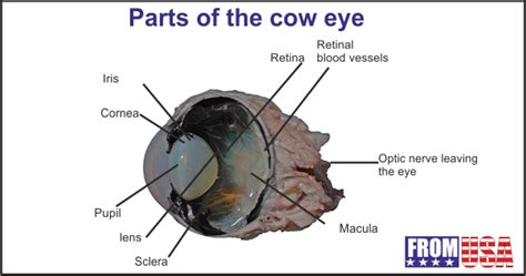
Table of Contents
- Label Gross Anatomy Of Cow Eye
- Table of Contents
- Labelled Gross Anatomy of a Cow Eye: A Comprehensive Guide
- External Anatomy of the Cow Eye
- 1. The Eyeball (Bulbus Oculi):
- 2. Conjunctiva:
- 3. Sclera:
- 4. Cornea:
- 5. Iris:
- 6. Pupil:
- 7. Eyelids (Palpebrae):
- 8. Nictitating Membrane (Third Eyelid):
- Internal Anatomy of the Cow Eye
- 1. Lens:
- 2. Ciliary Body:
- 3. Aqueous Humor:
- 4. Vitreous Humor:
- 5. Retina:
- 6. Choroid:
- 7. Optic Nerve:
- 8. Tapetum Lucidum:
- Practical Applications of Labeling the Cow Eye
- Potential Challenges and Considerations
- Conclusion
- Latest Posts
- Latest Posts
- Related Post
Labelled Gross Anatomy of a Cow Eye: A Comprehensive Guide
The bovine eye, remarkably similar in structure to the human eye, provides an excellent model for studying mammalian ophthalmic anatomy. Dissecting and labeling a cow eye offers a hands-on learning experience, allowing for a detailed understanding of its intricate components. This comprehensive guide will delve into the gross anatomy of the cow eye, providing a detailed description of each structure and its function. We'll explore the external features, the internal components, and their interconnectedness, emphasizing the practical application of labeling these structures accurately.
External Anatomy of the Cow Eye
Before delving into the internal structures, let's examine the external features visible before dissection. These external features are crucial for understanding the overall protection and function of the eye.
1. The Eyeball (Bulbus Oculi):
This is the spherical structure itself, containing the majority of the eye's components. Its size and shape are key to its function in focusing light and protecting the delicate internal structures.
2. Conjunctiva:
This thin, transparent mucous membrane lines the inner surface of the eyelids and covers the sclera (the white part of the eye). It's crucial for lubrication and protection of the eye's surface. Note: When dissecting, observe its delicate nature and careful handling is required to avoid tearing.
3. Sclera:
The tough, white outer layer of the eyeball. It provides structural support and protection for the more delicate internal structures. The sclera's strong fibrous nature is essential for maintaining the eye's shape. Look for: The point where the sclera transitions to the cornea.
4. Cornea:
This transparent, dome-shaped structure at the front of the eye is crucial for focusing light. Its curvature refracts light rays, bending them towards the lens. Note: Its clarity is vital for visual acuity; any damage can significantly impair vision.
5. Iris:
The colored part of the eye surrounding the pupil. The iris contains muscles that control the size of the pupil, regulating the amount of light entering the eye. Observe: The variations in color and pattern amongst different cow eyes.
6. Pupil:
The black circular opening in the center of the iris through which light enters the eye. The pupil's diameter is dynamically adjusted by the iris to adapt to varying light conditions. Focus on: The pupillary light reflex—observe how the pupil constricts in response to bright light (if you have a light source available during the dissection).
7. Eyelids (Palpebrae):
These protective folds of skin cover and protect the eye. They help to keep the eye lubricated and clear of debris. Examine: The eyelashes and Meibomian glands within the eyelids, contributing to tear film production.
8. Nictitating Membrane (Third Eyelid):
A thin, transparent membrane found in the medial corner of the eye. While less developed than in some other animals, it aids in protecting the cornea and distributing lubricating fluid. Look for: Its location and its relatively translucent nature.
Internal Anatomy of the Cow Eye
Once the external features are identified, careful dissection reveals the intricate internal structures. This dissection process allows for detailed examination and understanding of the optical and neural components.
1. Lens:
A transparent, biconvex structure located behind the iris. The lens focuses light onto the retina, crucial for sharp vision. Its elasticity allows it to adjust its shape to focus on objects at different distances (accommodation). Note: Its delicate nature requires careful handling during dissection.
2. Ciliary Body:
The ring of tissue surrounding the lens. It contains the ciliary muscles that control the shape of the lens, enabling accommodation. The ciliary body also produces aqueous humor. Look for: Its connection to the lens and iris.
3. Aqueous Humor:
A clear, watery fluid filling the space between the cornea and the lens (anterior chamber). It provides nutrients to the cornea and lens and helps maintain intraocular pressure. Observe: Its clear, watery nature during dissection (it may have drained away during processing).
4. Vitreous Humor:
A clear, gel-like substance filling the space between the lens and the retina (posterior chamber). It helps maintain the shape of the eyeball and provides support for the retina. Note: Its consistency and its role in maintaining the eye's shape.
5. Retina:
The light-sensitive innermost layer of the eye lining the posterior chamber. It contains photoreceptor cells (rods and cones) that convert light into electrical signals. These signals are then transmitted to the brain via the optic nerve. Examine: The delicate nature of the retina and its layered structure (although detailed microscopic examination is beyond the scope of gross anatomy).
6. Choroid:
A highly vascular layer located between the sclera and the retina. It provides oxygen and nutrients to the outer layers of the retina. Look for: Its dark pigmentation, which absorbs scattered light and reduces glare within the eye.
7. Optic Nerve:
A bundle of nerve fibers that transmits visual information from the retina to the brain. Locate: The point where the optic nerve exits the eyeball (the optic disc or blind spot).
8. Tapetum Lucidum:
A reflective layer behind the retina found in many nocturnal animals, including cows. It reflects light back onto the retina, enhancing vision in low-light conditions. Observe: Its iridescent appearance and its contribution to the "eyeshine" effect visible in cows at night.
Practical Applications of Labeling the Cow Eye
Labeling a cow eye during dissection is not just a classroom exercise; it reinforces the understanding of complex anatomical relationships. Accurate labeling is critical for demonstrating a grasp of the spatial arrangement of these structures and their functional interconnections.
Tips for effective labeling:
- Use clear and concise labels: Avoid ambiguous terms. Use standard anatomical terminology.
- Precise placement: Ensure labels accurately reflect the location of each structure.
- Consistent style: Maintain a consistent font, size, and color for labels.
- Layered approach: Label external features first, then progressively label internal structures as you dissect.
- Use a diagram: A pre-drawn diagram is useful for planning and recording your observations.
Potential Challenges and Considerations
Dissection of a cow eye presents certain challenges. The structures are delicate, requiring careful handling to avoid damage. Furthermore, the precise identification of some structures may require practice and a strong understanding of anatomical terminology. Fresh eyes are generally preferred for optimal clarity and preservation of structures. If using preserved specimens, be mindful of potential artifacts or distortions.
Conclusion
The cow eye, with its striking similarity to the human eye, offers a unique opportunity to learn about mammalian ocular anatomy. Through careful dissection and accurate labeling, one can develop a comprehensive understanding of the eye's complex structure and the functional interplay of its various components. This knowledge is invaluable not only for students of biology and veterinary science but also for anyone interested in the wonders of the visual system. Remember, accurate labeling is a crucial step in effectively communicating your anatomical understanding. This guide should empower you to tackle the dissection and labeling process confidently, leading to a richer and more rewarding learning experience. Remember to always prioritize safety and follow appropriate dissection protocols.
Latest Posts
Latest Posts
Related Post
Thank you for visiting our website which covers about Label Gross Anatomy Of Cow Eye . We hope the information provided has been useful to you. Feel free to contact us if you have any questions or need further assistance. See you next time and don't miss to bookmark.
