Label The Circulatory System Answer Key
Onlines
Mar 06, 2025 · 5 min read
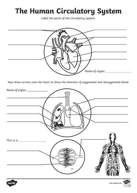
Table of Contents
Labeling the Circulatory System: A Comprehensive Guide with Answer Key
The circulatory system, also known as the cardiovascular system, is a vital network responsible for transporting blood, oxygen, nutrients, hormones, and other essential substances throughout the body. Understanding its intricate structure is crucial for anyone studying biology, anatomy, or related fields. This comprehensive guide will not only help you accurately label the circulatory system but also provide a deep understanding of its components and functions. We'll cover the key elements, their roles, and finally, provide an answer key to solidify your knowledge.
Major Components of the Circulatory System
The circulatory system is broadly divided into two main circuits: the pulmonary circulation and the systemic circulation. Let's break down the key components within each:
1. The Heart: The Central Pump
The heart, a muscular organ roughly the size of a fist, is the powerhouse of the circulatory system. Its rhythmic contractions propel blood through the body. Key structures within the heart include:
- Right Atrium: Receives deoxygenated blood returning from the body.
- Right Ventricle: Pumps deoxygenated blood to the lungs.
- Left Atrium: Receives oxygenated blood from the lungs.
- Left Ventricle: Pumps oxygenated blood to the rest of the body.
- Aorta: The largest artery in the body, carrying oxygenated blood from the left ventricle.
- Pulmonary Artery: Carries deoxygenated blood from the right ventricle to the lungs.
- Pulmonary Veins: Carry oxygenated blood from the lungs to the left atrium.
- Superior Vena Cava: Returns deoxygenated blood from the upper body to the right atrium.
- Inferior Vena Cava: Returns deoxygenated blood from the lower body to the right atrium.
- Tricuspid Valve: Located between the right atrium and right ventricle.
- Pulmonary Valve: Located between the right ventricle and pulmonary artery.
- Mitral (Bicuspid) Valve: Located between the left atrium and left ventricle.
- Aortic Valve: Located between the left ventricle and the aorta.
- Sinoatrial (SA) Node: The heart's natural pacemaker, initiating the heartbeat.
- Atrioventricular (AV) Node: Delays the electrical impulse, allowing the atria to fully contract before the ventricles.
- Bundle of His: Conducts the electrical impulse to the ventricles.
- Purkinje Fibers: Distribute the electrical impulse throughout the ventricles, causing them to contract.
2. Blood Vessels: The Transport Network
Blood vessels form a vast network that carries blood throughout the body. They are categorized into three main types:
- Arteries: Carry oxygenated blood away from the heart (except for the pulmonary artery). They have thick, elastic walls to withstand the high pressure of blood pumped from the heart. Arteries branch into smaller arterioles.
- Veins: Carry deoxygenated blood back to the heart (except for the pulmonary veins). They have thinner walls than arteries and often contain valves to prevent backflow of blood. Smaller venules merge to form veins.
- Capillaries: Microscopic vessels connecting arterioles and venules. Their thin walls allow for the exchange of gases, nutrients, and waste products between blood and tissues.
3. Blood: The Transport Medium
Blood is a specialized connective tissue that plays a crucial role in transporting various substances throughout the body. Its main components include:
- Red Blood Cells (Erythrocytes): Carry oxygen bound to hemoglobin.
- White Blood Cells (Leukocytes): Part of the immune system, fighting infections.
- Platelets (Thrombocytes): Involved in blood clotting.
- Plasma: The liquid component of blood, containing water, proteins, hormones, and other dissolved substances.
Pulmonary Circulation: The Lung Circuit
Pulmonary circulation involves the movement of blood between the heart and the lungs. Deoxygenated blood from the body enters the right atrium, passes through the tricuspid valve into the right ventricle, and is then pumped through the pulmonary artery to the lungs. In the lungs, carbon dioxide is exchanged for oxygen. Oxygenated blood then returns to the heart via the pulmonary veins, entering the left atrium.
Systemic Circulation: The Body Circuit
Systemic circulation involves the movement of blood between the heart and the rest of the body. Oxygenated blood from the left atrium passes through the mitral valve into the left ventricle. The left ventricle pumps this blood through the aorta, the body's largest artery, to various organs and tissues. After delivering oxygen and nutrients, and picking up carbon dioxide and waste products, deoxygenated blood returns to the heart through the vena cavae.
Labeling Exercise and Answer Key
Now, let's test your understanding with a labeling exercise. Below is a simplified diagram of the circulatory system. Identify the labeled structures. (Note: A visual diagram would be included here in a real document. For the purpose of this text-based response, I'll provide a textual representation, which is less effective for this type of exercise.)
(Textual Representation of a Diagram - Imagine a heart with major vessels)
- (Structure A: Right Atrium)
- (Structure B: Right Ventricle)
- (Structure C: Left Atrium)
- (Structure D: Left Ventricle)
- (Structure E: Aorta)
- (Structure F: Pulmonary Artery)
- (Structure G: Pulmonary Veins)
- (Structure H: Superior Vena Cava)
- (Structure I: Inferior Vena Cava)
Answer Key:
- Structure A: Right Atrium
- Structure B: Right Ventricle
- Structure C: Left Atrium
- Structure D: Left Ventricle
- Structure E: Aorta
- Structure F: Pulmonary Artery
- Structure G: Pulmonary Veins
- Structure H: Superior Vena Cava
- Structure I: Inferior Vena Cava
Advanced Concepts and Further Exploration
This guide has provided a foundational understanding of the circulatory system. For a more in-depth understanding, consider exploring these advanced concepts:
- Cardiac Cycle: The sequence of events in a single heartbeat.
- Blood Pressure: The force exerted by blood against the walls of blood vessels.
- Regulation of Heart Rate: The mechanisms that control the speed of the heartbeat.
- Blood Types and Transfusions: The different blood groups and their compatibility.
- Hematopoiesis: The process of blood cell formation.
- Diseases of the Circulatory System: Conditions like heart disease, stroke, and hypertension.
By studying these aspects, you'll gain a comprehensive grasp of this intricate system and its vital role in maintaining human health. Remember to consult reputable textbooks and online resources for a more detailed and visually enhanced learning experience. This article serves as a starting point for your exploration of this fascinating topic. Understanding the circulatory system is a key component of understanding the human body as a whole. The more you learn, the more you will appreciate the complexity and efficiency of this remarkable system.
Latest Posts
Latest Posts
-
Which Of The Following Can Be Controlled By Copyright
Mar 06, 2025
-
Which Sentence Correctly Uses Parallel Structure
Mar 06, 2025
-
My Fathers Eyes My Mothers Rage Pdf Free Download
Mar 06, 2025
-
What Macromolecules Provide Energy For Lions And Elephants
Mar 06, 2025
-
List The Six Principles Associated With Bond Pricing Relationships
Mar 06, 2025
Related Post
Thank you for visiting our website which covers about Label The Circulatory System Answer Key . We hope the information provided has been useful to you. Feel free to contact us if you have any questions or need further assistance. See you next time and don't miss to bookmark.
