Label The Transmission Electron Micrograph Of The Nucleus
Onlines
Apr 02, 2025 · 5 min read
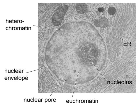
Table of Contents
- Label The Transmission Electron Micrograph Of The Nucleus
- Table of Contents
- Labeling a Transmission Electron Micrograph (TEM) of the Nucleus: A Comprehensive Guide
- Key Structures within the Nucleus: A Visual Inventory
- 1. Nuclear Envelope: The Protective Barrier
- 2. Nucleolus: The Ribosome Factory
- 3. Chromatin: The Genetic Material
- 4. Nucleoplasm: The Nuclear Matrix
- Step-by-Step Guide to Labeling Your TEM
- Advanced Considerations: Beyond the Basics
- Interpreting the Image: Beyond Simple Labeling
- Conclusion: Mastering TEM Interpretation
- Latest Posts
- Latest Posts
- Related Post
Labeling a Transmission Electron Micrograph (TEM) of the Nucleus: A Comprehensive Guide
The nucleus, the control center of eukaryotic cells, is a complex organelle teeming with intricate structures. Understanding its ultrastructure requires careful examination of transmission electron micrographs (TEMs). This detailed guide will walk you through the process of accurately labeling a TEM of a nucleus, covering key structures and their distinguishing features. We'll delve into the intricacies of nuclear morphology, highlighting the importance of proper identification for accurate cellular interpretation.
Key Structures within the Nucleus: A Visual Inventory
Before we dive into labeling, let's review the essential components visible in a typical TEM of a nucleus:
1. Nuclear Envelope: The Protective Barrier
-
Outer Membrane: This membrane is continuous with the endoplasmic reticulum (ER) and often studded with ribosomes, reflecting its role in protein synthesis. Label this clearly on your micrograph.
-
Inner Membrane: This membrane lines the nucleoplasm and is associated with the nuclear lamina, a protein meshwork providing structural support. Remember to identify this layer.
-
Nuclear Pores: These complex structures regulate the transport of molecules between the nucleus and cytoplasm. They appear as distinct pores or interruptions in the nuclear envelope. Highlight these crucial structures in your labeling. Note their size and distribution.
2. Nucleolus: The Ribosome Factory
The nucleolus is a prominent, electron-dense region within the nucleus. Its appearance can vary depending on the cell's metabolic activity. It's responsible for ribosome biogenesis. Make sure to clearly mark the nucleolus on your diagram. Look for its characteristic granular and fibrillar regions.
3. Chromatin: The Genetic Material
Chromatin, composed of DNA and associated proteins, is the genetic material of the cell. In TEMs, it appears as either:
-
Euchromatin: Less condensed, lighter-staining regions representing actively transcribed genes. Differentiate this from heterochromatin in your labeling.
-
Heterochromatin: More condensed, darker-staining regions representing transcriptionally inactive DNA. Clearly label the areas of heterochromatin. Observe its distribution – often concentrated at the nuclear periphery.
4. Nucleoplasm: The Nuclear Matrix
The nucleoplasm is the gel-like substance filling the nuclear interior. It’s not a visually distinct structure like others, but it's essential to understand its role in nuclear organization and function. While you might not explicitly label "nucleoplasm," its presence is implied by the space occupied by the other labeled structures.
Step-by-Step Guide to Labeling Your TEM
Now, let's walk through the process of labeling a TEM of a nucleus, focusing on accuracy and clarity:
-
Obtain a high-quality image: Start with a clear, well-resolved TEM image. Low resolution or artifacts will hinder accurate labeling.
-
Identify the nuclear envelope: The nuclear envelope is the outermost boundary of the nucleus. It's relatively easy to identify due to its double-membrane structure and presence of nuclear pores. Label it clearly as "Nuclear Envelope."
-
Locate the nucleolus: Look for the electron-dense, usually round or ovoid structure within the nucleus. This is the nucleolus. Label it "Nucleolus."
-
Differentiate euchromatin and heterochromatin: Observe the differences in staining intensity within the nuclear interior. Lighter regions represent euchromatin, while darker regions represent heterochromatin. Label them accordingly.
-
Mark nuclear pores: These appear as distinct interruptions in the nuclear envelope. Label at least a few of these "Nuclear Pores" to demonstrate your understanding of their presence.
-
Use consistent and clear labeling: Employ clear labels, using a consistent font and size. Avoid overlapping labels and ensure they are close enough to the corresponding structure.
-
Consider adding a scale bar: Include a scale bar to indicate the magnification of the micrograph. This is crucial for providing context and allowing readers to judge the size of the structures.
-
Create a legend: If necessary, include a legend providing a key to the labels used. This is particularly helpful if you’re using abbreviations or color-coding.
Advanced Considerations: Beyond the Basics
Once you’re comfortable labeling the basic structures, consider these advanced elements:
-
Nuclear matrix: While not directly visible, understanding the nuclear matrix's role in organizing chromatin and other nuclear components enhances your interpretation. You might indicate its presence conceptually, even if it's not explicitly labeled.
-
Specific Chromatin Domains: Advanced TEM techniques might reveal specific chromatin domains associated with particular genes or regulatory elements. Labeling these depends on the image's resolution and the specific technique used to prepare the sample.
-
Nuclear bodies: Various nuclear bodies, including Cajal bodies and PML bodies, might be visible depending on the cell type and its activity. Their labeling would depend on their presence in the image and your level of expertise.
-
Nuclear lamina: While not always clearly visible in every TEM, its presence is implied by the structural integrity of the nuclear envelope. If visible, it should be clearly labeled.
Interpreting the Image: Beyond Simple Labeling
Correct labeling is only one part of interpreting a TEM of a nucleus. Consider these points:
-
Cell type: The appearance of the nucleus will vary depending on the cell type. For example, neurons typically have a larger and more prominent nucleolus than other cells.
-
Cell cycle: The organization of chromatin changes throughout the cell cycle. A cell in mitosis will show highly condensed chromosomes, whereas a cell in interphase will show more dispersed chromatin.
-
Cellular activity: The relative proportions of euchromatin and heterochromatin can reflect the cell's level of transcriptional activity. A cell actively synthesizing proteins will tend to have more euchromatin.
-
Pathological conditions: Abnormalities in nuclear structure can indicate cellular dysfunction or disease. For example, altered nuclear shape or size can be indicative of cancerous cells.
Conclusion: Mastering TEM Interpretation
Accurate labeling of a transmission electron micrograph of the nucleus requires a solid understanding of its components and their functions. By following the steps outlined in this guide and paying close attention to the details of the image, you can master the art of TEM interpretation and gain valuable insights into the cellular world. Remember, accurate labeling is not just about identifying structures; it's about understanding their significance within the context of cellular biology and function. Practice analyzing different TEM images, and soon you’ll be confidently interpreting the intricate structures of the nucleus. Continuously updating your knowledge of cellular biology will further enhance your ability to accurately label and interpret TEM images of the nucleus.
Latest Posts
Latest Posts
-
Natural Selection Color By Number Answer Key
Apr 07, 2025
-
Which Situation Best Reflects The Concept Of Free Enterprise
Apr 07, 2025
-
2 09 Unit Test Radicals And Complex Numbers Part 1
Apr 07, 2025
-
Experiment 5 The Importance Of Cell Cycle Control
Apr 07, 2025
-
2 08 Quiz Rhetoric Develops Purpose And Viewpoint
Apr 07, 2025
Related Post
Thank you for visiting our website which covers about Label The Transmission Electron Micrograph Of The Nucleus . We hope the information provided has been useful to you. Feel free to contact us if you have any questions or need further assistance. See you next time and don't miss to bookmark.