Pal Cadaver Appendicular Skeleton Upper Limb Lab Practical Question 3
Onlines
Mar 18, 2025 · 6 min read
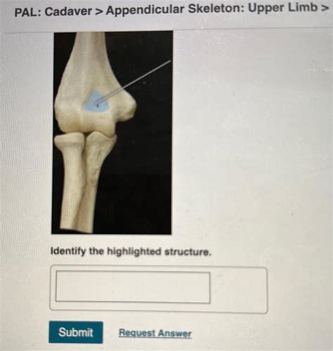
Table of Contents
Pal Cadaver Appendicular Skeleton Upper Limb Lab Practical Question 3: A Comprehensive Guide
This article delves into a common practical question encountered in anatomy labs: the examination of the upper limb appendicular skeleton using a pal cadaver. We'll cover key anatomical structures, practical dissection tips, and potential questions to expect during a lab practical. This comprehensive guide aims to equip students with the knowledge and confidence to excel in their anatomical studies.
Understanding the Appendicular Skeleton of the Upper Limb
The appendicular skeleton of the upper limb comprises the bones that form the arm, forearm, and hand. Its intricate structure allows for a wide range of movement and dexterity. A thorough understanding requires familiarity with each bone's individual features and their articulation (joint formation) with neighboring bones.
The Major Bones:
-
Clavicle (Collarbone): A long, S-shaped bone connecting the sternum (breastbone) to the scapula (shoulder blade). Note its medial (sternal) and lateral (acromial) ends. Palpation of the clavicle is relatively easy on a living subject. Look for its prominent curvature on the cadaver.
-
Scapula (Shoulder Blade): A flat, triangular bone situated on the posterior thorax. Identify its important features: the acromion process (articulates with the clavicle), the coracoid process (serves as an attachment point for muscles), the glenoid cavity (socket for the humerus), and the spine of the scapula (a prominent ridge).
-
Humerus (Upper Arm Bone): The longest bone of the upper limb. Observe its proximal (shoulder) end, featuring the head (articulates with the glenoid cavity), the greater and lesser tubercles (muscle attachment sites), and the anatomical neck. The distal (elbow) end showcases the medial and lateral epicondyles, the capitulum (articulates with the radius), and the trochlea (articulates with the ulna).
-
Radius (Lateral Forearm Bone): Located on the lateral (thumb) side of the forearm. Observe its proximal head (articulates with the capitulum of the humerus and the radial notch of the ulna), the radial tuberosity (muscle attachment), and the distal end which articulates with the carpal bones of the wrist.
-
Ulna (Medial Forearm Bone): Situated on the medial (pinky finger) side of the forearm. Identify its proximal olecranon process (forms the point of the elbow), the trochlear notch (articulates with the trochlea of the humerus), the radial notch (articulates with the head of the radius), and the distal head of the ulna.
-
Carpal Bones (Wrist Bones): Eight small bones arranged in two rows. While memorizing individual carpal bones (scaphoid, lunate, triquetrum, pisiform, trapezium, trapezoid, capitate, hamate) can be challenging, focus on identifying the overall arrangement and their relationships with the radius and ulna proximally, and the metacarpals distally.
-
Metacarpal Bones (Palm Bones): Five long bones forming the palm of the hand. Note their numbering (I-V, starting from the thumb).
-
Phalanges (Finger Bones): Fourteen bones forming the fingers. Each finger (except the thumb) has three phalanges: proximal, middle, and distal. The thumb only has two: proximal and distal.
Practical Dissection and Examination of the Pal Cadaver
Examining a pal cadaver requires a delicate and respectful approach. Always follow the instructions provided by your instructor. Proper handling and hygiene practices are paramount.
Step-by-Step Approach:
-
Initial Observation: Begin by visually inspecting the entire upper limb. Note the overall position and arrangement of the bones.
-
Careful Removal of Superficial Tissues: Gently remove the superficial tissues (skin, subcutaneous fat) layer by layer. This will reveal the underlying muscles and then the bones. Use fine dissection instruments such as forceps and scalpel to avoid damaging the bones.
-
Identifying Key Landmarks: Once the bones are exposed, carefully identify the key bony landmarks mentioned above. Using anatomical models and atlases in conjunction with the cadaver can significantly improve identification accuracy.
-
Articulations and Movements: Gently manipulate the joints (articulations) to understand their range of motion. Observe how the bones fit together.
-
Bone Features and Characteristics: Closely examine the texture, shape, and size of each bone. Look for any abnormalities or variations from the typical anatomical presentation.
-
Detailed Examination of Specific Areas: Pay close attention to areas that are frequently tested, such as the elbow joint (articulation between the humerus, radius, and ulna), the wrist joint (articulation between the radius, ulna, and carpals), and the hand.
-
Documentation: Take detailed notes and, if allowed, photographs to document your observations. This aids retention and understanding.
Potential Lab Practical Questions
Lab practical exams assess your understanding of the anatomy through direct observation and manipulation. Here are some potential questions related to the pal cadaver upper limb appendicular skeleton:
- Identify the bones of the upper limb. This is a fundamental question testing basic knowledge.
- Identify specific bony landmarks on the humerus, radius, and ulna. This requires detailed knowledge of bone features.
- Describe the articulations of the elbow joint. Understanding the joint mechanics is crucial.
- Compare and contrast the radius and ulna. This probes your understanding of anatomical differences.
- Explain the arrangement and function of the carpal bones. This focuses on the wrist's intricate structure.
- Demonstrate the range of motion at the shoulder, elbow, and wrist joints. This is a practical demonstration of your understanding.
- Identify the bones contributing to the formation of the wrist and hand. This requires a thorough understanding of the distal upper limb.
- Differentiate between the proximal and distal ends of the humerus and ulna. This assesses your detailed observation skills.
- Explain the significance of specific bony landmarks in muscle attachment. This integrates muscular anatomy with skeletal anatomy.
- Identify any anomalies or variations in bone structure. This tests your ability to recognize differences from the norm.
Advanced Considerations and Further Learning
While the basic anatomical structures are essential, mastering the upper limb’s intricacies demands deeper engagement.
Neurovascular Structures:
Understanding the relationship between the bones and the nerves and blood vessels that traverse the upper limb is crucial for clinical practice. Consider the brachial plexus and its branches, as well as the major arteries and veins supplying the arm and hand.
Clinical Correlations:
Relating the anatomical structures to common injuries and pathologies is vital. For example, understanding the location of the ulnar nerve in relation to the medial epicondyle is crucial for diagnosing cubital tunnel syndrome. Consider fractures, dislocations, and other injuries to further your knowledge.
Imaging Techniques:
Familiarize yourself with radiographic images of the upper limb. Learning to interpret X-rays and other imaging modalities enhances the practical application of your anatomical knowledge.
Conclusion
Mastering the anatomy of the upper limb appendicular skeleton using a pal cadaver requires meticulous observation, careful dissection, and a thorough understanding of the anatomical terminology. This guide provides a comprehensive framework for approaching your lab practical, emphasizing detailed examination and clinical correlations. Through dedicated study and practical application, you can build a strong foundation in human anatomy and achieve success in your academic pursuits. Remember to always approach cadaveric dissection with respect and adhere to all safety protocols and guidelines provided by your instructor. Good luck with your studies!
Latest Posts
Latest Posts
-
Pal Cadaver Appendicular Skeleton Joints Lab Practical Question 10
Mar 18, 2025
-
Huipil Es Una De Las Ciudades Mas Importantes De Guatemala
Mar 18, 2025
-
The Decision Making Process In Driving Is Known As
Mar 18, 2025
-
A Wall Of Fire Rising Summary
Mar 18, 2025
-
Highway Hypnosis Is Related To
Mar 18, 2025
Related Post
Thank you for visiting our website which covers about Pal Cadaver Appendicular Skeleton Upper Limb Lab Practical Question 3 . We hope the information provided has been useful to you. Feel free to contact us if you have any questions or need further assistance. See you next time and don't miss to bookmark.
